Explore
 Validate
 Learn
Learn Western blot
Antibody data
- Antibody Data
- Antigen structure
- References [1]
- Comments [0]
- Validations
- Western blot [2]
- Immunocytochemistry [1]
- Immunohistochemistry [8]
- Flow cytometry [2]
Submit
Validation data
Reference
Comment
Report error
- Product number
- TA503471 - Provider product page

- Provider
- OriGene
- Proper citation
- OriGene Cat#TA503471, RRID:AB_11125900
- Product name
- PDE4B mouse monoclonal antibody, clone OTI1D12 (formerly 1D12)
- Antibody type
- Monoclonal
- Description
- PDE4B mouse monoclonal antibody, clone OTI1D12 (formerly 1D12)
- Host
- Mouse
- Conjugate
- Unconjugated
- Epitope
- PDE4B
- Isotype
- IgG
- Antibody clone number
- OTI1D12
- Vial size
- 100 µl
- Concentration
- 0.62 mg/ml
Submitted references AMPK antagonizes hepatic glucagon-stimulated cyclic AMP signalling via phosphorylation-induced activation of cyclic nucleotide phosphodiesterase 4B.
Johanns M, Lai YC, Hsu MF, Jacobs R, Vertommen D, Van Sande J, Dumont JE, Woods A, Carling D, Hue L, Viollet B, Foretz M, Rider MH
Nature communications 2016 Mar 8;7:10856
Nature communications 2016 Mar 8;7:10856
No comments: Submit comment
Supportive validation
- Submitted by
- OriGene (provider)
- Main image
- Experimental details
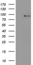
- HEK293T cells were transfected with the pCMV6-ENTRY control (Left lane) or pCMV6-ENTRY PDE4B (RC211956, Right lane) cDNA for 48 hrs and lysed. Equivalent amounts of cell lysates (5 ug per lane) were separated by SDS-PAGE and immunoblotted with anti-PDE4B.
- Validation comment
- WB
- Submitted by
- OriGene (provider)
- Main image
- Experimental details
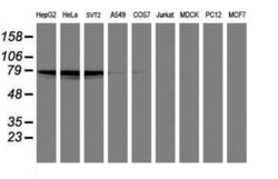
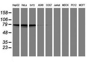
- Western blot analysis of extracts (35ug) from 9 different cell lines by using anti-PDE4B monoclonal antibody (HepG2: human; HeLa: human; SVT2: mouse; A549: human; COS7: monkey; Jurkat: human; MDCK: canine; PC12: rat; MCF7: human).
- Validation comment
- WB
Supportive validation
- Submitted by
- OriGene (provider)
- Main image

- Experimental details
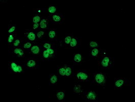
- Anti-PDE4B mouse monoclonal antibody (TA503471) immunofluorescent staining of COS7 cells transiently transfected by pCMV6-ENTRY PDE4B(RC211956).
- Validation comment
- IF
Supportive validation
- Submitted by
- OriGene (provider)
- Main image
- Experimental details
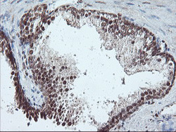
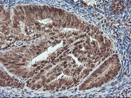
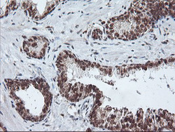
- Immunohistochemical staining of paraffin-embedded Human prostate tissue within the normal limits using anti-PDE4B mouse monoclonal antibody. (Heat-induced epitope retrieval by 10mM citric buffer, pH6.0, 100C for 10min, TA503471)
- Validation comment
- IHC
- Submitted by
- OriGene (provider)
- Main image

- Experimental details
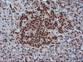
- Immunohistochemical staining of paraffin-embedded Adenocarcinoma of Human endometrium tissue using anti-PDE4B mouse monoclonal antibody. (Heat-induced epitope retrieval by 10mM citric buffer, pH6.0, 100C for 10min, TA503471)
- Validation comment
- IHC
- Submitted by
- OriGene (provider)
- Main image

- Experimental details
- Immunohistochemical staining of paraffin-embedded Human pancreas tissue within the normal limits using anti-PDE4B mouse monoclonal antibody. (Heat-induced epitope retrieval by 10mM citric buffer, pH6.0, 100C for 10min, TA503471)
- Validation comment
- IHC
- Submitted by
- OriGene (provider)
- Main image
- Experimental details
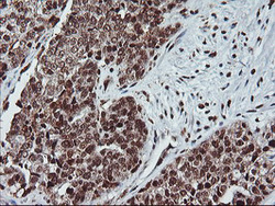
- Immunohistochemical staining of paraffin-embedded Adenocarcinoma of Human ovary tissue using anti-PDE4B mouse monoclonal antibody. (Heat-induced epitope retrieval by 10mM citric buffer, pH6.0, 100C for 10min, TA503471)
- Validation comment
- IHC
- Submitted by
- OriGene (provider)
- Main image
- Experimental details
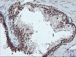
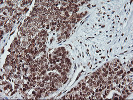
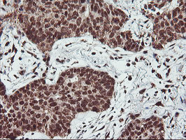
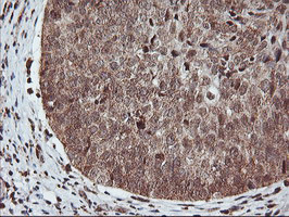
- Immunohistochemical staining of paraffin-embedded Carcinoma of Human prostate tissue using anti-PDE4B mouse monoclonal antibody. (Heat-induced epitope retrieval by 10mM citric buffer, pH6.0, 100C for 10min, TA503471)
- Validation comment
- IHC

- Submitted by
- OriGene (provider)
- Main image

- Experimental details
- Immunohistochemical staining of paraffin-embedded Carcinoma of Human lung tissue using anti-PDE4B mouse monoclonal antibody. (Heat-induced epitope retrieval by 10mM citric buffer, pH6.0, 100C for 10min, TA503471)
- Validation comment
- IHC
- Submitted by
- OriGene (provider)
- Main image
- Experimental details
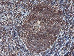
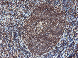
- Immunohistochemical staining of paraffin-embedded Human tonsil within the normal limits using anti-PDE4B mouse monoclonal antibody. (Heat-induced epitope retrieval by 10mM citric buffer, pH6.0, 100C for 10min, TA503471)
- Validation comment
- IHC
- Submitted by
- OriGene (provider)
- Main image

- Experimental details
- Immunohistochemical staining of paraffin-embedded Carcinoma of Human bladder tissue using anti-PDE4B mouse monoclonal antibody. (Heat-induced epitope retrieval by 10mM citric buffer, pH6.0, 100C for 10min, TA503471)
- Validation comment
- IHC
Supportive validation
- Submitted by
- OriGene (provider)
- Main image
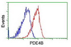
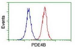
- Experimental details
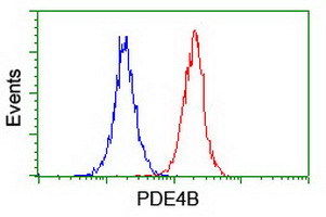
- Flow cytometric Analysis of Hela cells, using anti-PDE4B antibody(TA503471),(Red), compared to a nonspecific negative control antibody,(Blue).
- Validation comment
- FC
- Submitted by
- OriGene (provider)
- Main image
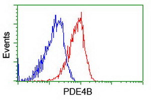
- Experimental details
- Flow cytometric Analysis of Jurkat cells, using anti-PDE4B antibody(TA503471),(Red), compared to a nonspecific negative control antibody,(Blue).
- Validation comment
- FC