 Explore
 Validate
 Learn
Learn Western blot
Antibody data
- Antibody Data
- Antigen structure
- References [3]
- Comments [0]
- Validations
- Western blot [1]
- Other assay [3]
Submit
Validation data
Reference
Comment
Report error
- Product number
- PA5-17624 - Provider product page

- Provider
- Invitrogen Antibodies
- Product name
- MYL9 Polyclonal Antibody
- Antibody type
- Polyclonal
- Antigen
- Synthetic peptide
- Description
- It is not recommended to aliquot this antibody. This antibody is not cross-reactive with the cardiac isoform of Myosin Light Chain 2, or Myosin Essential Light Chain.
- Reactivity
- Human, Mouse, Rat
- Host
- Rabbit
- Isotype
- IgG
- Vial size
- 100 µL
- Concentration
- 19 µg/mL
- Storage
- -20°C
Submitted references ADF and cofilin-1 collaborate to promote cortical actin flow and the leader bleb-based migration of confined cells.
Rnd3 regulates lung cancer cell proliferation through notch signaling.
Rnd3 haploinsufficient mice are predisposed to hemodynamic stress and develop apoptotic cardiomyopathy with heart failure.
Ullo MF, Logue JS
eLife 2021 Jun 25;10
eLife 2021 Jun 25;10
Rnd3 regulates lung cancer cell proliferation through notch signaling.
Tang Y, Hu C, Yang H, Cao L, Li Y, Deng P, Huang L
PloS one 2014;9(11):e111897
PloS one 2014;9(11):e111897
Rnd3 haploinsufficient mice are predisposed to hemodynamic stress and develop apoptotic cardiomyopathy with heart failure.
Yue X, Yang X, Lin X, Yang T, Yi X, Dai Y, Guo J, Li T, Shi J, Wei L, Fan GC, Chen C, Chang J
Cell death & disease 2014 Jun 5;5(6):e1284
Cell death & disease 2014 Jun 5;5(6):e1284
No comments: Submit comment
Supportive validation
- Submitted by
- Invitrogen Antibodies (provider)
- Main image

- Experimental details
- Western blot was performed using Anti-MYL9 Polyclonal Antibody (Product # PA5-17624) and a 20 kDa band corresponding to Myosin regulatory light polypeptide 9 was observed across cell lines and tissue extract tested and not in Daudi, Mouse Skeletal Muscle and Mouse Liver which is reported to be negative. Whole cell extracts (30 µg lysate) of HeLa (Lane 1), Caco-2 (Lane 2), U-2 OS (Lane 3), Jurkat (Lane 4), MDA-MB-231 (Lane 5), C2C12 (Lane 6), Daudi (Lane 7) and tissue extracts Mouse Skeletal Muscle (Lane 8), Mouse Liver (Lane 9) and Mouse Urinary Bladder (Lane 10) were electrophoresed using NuPAGE™ 12% Bis-Tris Protein Gel (Product # NP0342BOX). Resolved proteins were then transferred onto a Nitrocellulose membrane (Product # IB23001) by iBlot® 2 Dry Blotting System (Product # IB21001). The blot was probed with the primary antibody (1:1000 dilution) and detected by chemiluminescence with Goat anti-Rabbit IgG (H+L) Superclonal™ Recombinant Secondary Antibody, HRP (Product # A27036, 1:4000 dilution) using the iBright FL 1000 (Product # A32752). Chemiluminescent detection was performed using Novex® ECL Chemiluminescent Substrate Reagent Kit (Product # WP20005).

Supportive validation
- Submitted by
- Invitrogen Antibodies (provider)
- Main image
- Experimental details
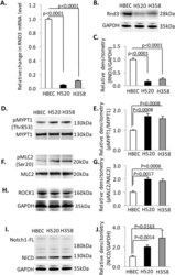
- Figure 1 Rnd3 is down-regulated in non-small lung cancer cell lines, H520 and H358. ( A ) Rnd3 mRNA detected by qRT-PCR is down-regulated in H358 and H520 compared to HBEC. ( B ) Rnd3 protein expression levels in cells by western blot. ( C ) Densitometry quantification of western band intensity in B. ( D ), ( F ), ( H ) & ( I ) A western blot to detect phosphorylated MYPT1, phosphorylated MLC2, ROCK1 and NICD in cells. ( E ), ( G ) & ( J ) Densitometry quantification of western band intensity showed up-regulation of Rho Kinase activity and NICD expression in H358 and H520 cells compared to HBEC. Western blots were quantified from three independent experimental repeats. BrdU-positive cells were quantified from 8 images taken from four slides. Data represent means +- S.D.

- Submitted by
- Invitrogen Antibodies (provider)
- Main image

- Experimental details
- Figure 3 Reintroduction of Rnd3 corrects Rho and Notch signaling in H520 and H358 cells. ( A ) & ( B ) Decreased NICD expression in H520-Rnd3 cells compared to H520 cells. ( C ) & ( D ) Decreased Rho Kinase activity in H520-Rnd3 cells compared to H520 cells detected by antibodies specific for pMYPT1 and pMLC2. ( E ) & ( F ) Decreased NICD expression in H358-Rnd3 cells compared to H358 cells. ( G ) & ( H ) Decreased Rho Kinase activity in H358-Rnd3 cells compared to H358 cells detected by antibodies specific for pMYPT1 and pMLC2. Western blots were quantified from three independent experimental repeats. Data represent means +- S.D.

- Submitted by
- Invitrogen Antibodies (provider)
- Main image

- Experimental details
- Figure 3 Complete deletion of Rnd3 gene resulted in severe apoptotic cardiomyopathy along with the elevation of Rho kinase activity in the mouse hearts. ( a ) A significant amount of TUNEL-positive cells were displayed in E10.5 Rnd3- hearts. The arrows indicate TUNEL-positive cells (green) overlapped with nucleus counter-staining. Cardiomyocytes were visualized by red fluorescent staining for F-actin. NS indicates nonspecific staining. ( b ) Quantification of the TUNEL-positive cells in hearts. ( c ) Elevated caspase-3 activity was detected in Rnd3- hearts by immunoblot analyses and quantified by immunoblot densitometry analyses. ( d ) Elevated Rho kinase activity was detected in Rnd3- hearts by immunoblotting for two Rho kinase substrates, MYPT1 and MLC2. The immunoblot densitometry analyses were quantified. Statistical significance was determined by unpaired, two-tailed Student's t -test. Data are means+-S.D. TUNEL, terminal deoxynucleotidyl transferase dUTP nick end labeling; MLC2, myosin light chain 2. The numbers in the columns represent the number of mice in each group
