Explore
 Validate
 Learn
Learn Western blot
Antibody data
- Antibody Data
- Antigen structure
- References [10]
- Comments [0]
- Validations
- Western blot [1]
- Immunocytochemistry [1]
- Other assay [4]
Submit
Validation data
Reference
Comment
Report error
- Product number
- PA5-17727 - Provider product page

- Provider
- Invitrogen Antibodies
- Product name
- Phospho-MYL9 (Thr18, Ser19) Polyclonal Antibody
- Antibody type
- Polyclonal
- Antigen
- Synthetic peptide
- Description
- It is not recommended to aliquot this antibody. This antibody is not cross-reactive with the cardiac isoform of myosin light chain 2.
- Reactivity
- Human, Mouse
- Host
- Rabbit
- Isotype
- IgG
- Vial size
- 100 μL
- Concentration
- 20 μg/mL
- Storage
- -20°C
Submitted references ADF and cofilin-1 collaborate to promote cortical actin flow and the leader bleb-based migration of confined cells.
Formin Activity and mDia1 Contribute to Maintain Axon Initial Segment Composition and Structure.
Factor quinolinone inhibitors alter cell morphology and motility by destabilizing interphase microtubules.
HIFα independent mechanisms in renal carcinoma cells modulate divergent outcomes in fibronectin assembly mediated by hypoxia and CoCl(2).
Histone deacetylase inhibitor CG200745 ameliorates high-fat diet-induced hypertension via inhibition of angiotensin II production.
17‑AAG synergizes with Belinostat to exhibit a negative effect on the proliferation and invasion of MDA‑MB‑231 breast cancer cells.
A two-phase response of endothelial cells to hydrostatic pressure.
Rnd3 regulates lung cancer cell proliferation through notch signaling.
Rnd3 haploinsufficient mice are predisposed to hemodynamic stress and develop apoptotic cardiomyopathy with heart failure.
New insights into myosin phosphorylation during cyclic nucleotide-mediated smooth muscle relaxation.
Ullo MF, Logue JS
eLife 2021 Jun 25;10
eLife 2021 Jun 25;10
Formin Activity and mDia1 Contribute to Maintain Axon Initial Segment Composition and Structure.
Zhang W, Ciorraga M, Mendez P, Retana D, Boumedine-Guignon N, Achón B, Russier M, Debanne D, Garrido JJ
Molecular neurobiology 2021 Dec;58(12):6153-6169
Molecular neurobiology 2021 Dec;58(12):6153-6169
Factor quinolinone inhibitors alter cell morphology and motility by destabilizing interphase microtubules.
Stoiber P, Scribani Rossi P, Pokharel N, Germany JL, York EA, Schaus SE, Hansen U
Scientific reports 2021 Dec 7;11(1):23564
Scientific reports 2021 Dec 7;11(1):23564
HIFα independent mechanisms in renal carcinoma cells modulate divergent outcomes in fibronectin assembly mediated by hypoxia and CoCl(2).
Magdaleno C, Dixon L, Rajasekaran N, Varadaraj A
Scientific reports 2020 Oct 29;10(1):18560
Scientific reports 2020 Oct 29;10(1):18560
Histone deacetylase inhibitor CG200745 ameliorates high-fat diet-induced hypertension via inhibition of angiotensin II production.
Yoon GE, Jung JK, Lee YH, Jang BC, In Kim J
Naunyn-Schmiedeberg's archives of pharmacology 2020 Mar;393(3):491-500
Naunyn-Schmiedeberg's archives of pharmacology 2020 Mar;393(3):491-500
17‑AAG synergizes with Belinostat to exhibit a negative effect on the proliferation and invasion of MDA‑MB‑231 breast cancer cells.
Zuo Y, Xu H, Chen Z, Xiong F, Zhang B, Chen K, Jiang H, Luo C, Zhang H
Oncology reports 2020 Jun;43(6):1928-1944
Oncology reports 2020 Jun;43(6):1928-1944
A two-phase response of endothelial cells to hydrostatic pressure.
Prystopiuk V, Fels B, Simon CS, Liashkovich I, Pasrednik D, Kronlage C, Wedlich-Söldner R, Oberleithner H, Fels J
Journal of cell science 2018 Jun 21;131(12)
Journal of cell science 2018 Jun 21;131(12)
Rnd3 regulates lung cancer cell proliferation through notch signaling.
Tang Y, Hu C, Yang H, Cao L, Li Y, Deng P, Huang L
PloS one 2014;9(11):e111897
PloS one 2014;9(11):e111897
Rnd3 haploinsufficient mice are predisposed to hemodynamic stress and develop apoptotic cardiomyopathy with heart failure.
Yue X, Yang X, Lin X, Yang T, Yi X, Dai Y, Guo J, Li T, Shi J, Wei L, Fan GC, Chen C, Chang J
Cell death & disease 2014 Jun 5;5(6):e1284
Cell death & disease 2014 Jun 5;5(6):e1284
New insights into myosin phosphorylation during cyclic nucleotide-mediated smooth muscle relaxation.
Puetz S, Schroeter MM, Piechura H, Reimann L, Hunger MS, Lubomirov LT, Metzler D, Warscheid B, Pfitzer G
Journal of muscle research and cell motility 2012 Dec;33(6):471-83
Journal of muscle research and cell motility 2012 Dec;33(6):471-83
No comments: Submit comment
Supportive validation
- Submitted by
- Invitrogen Antibodies (provider)
- Main image

- Experimental details
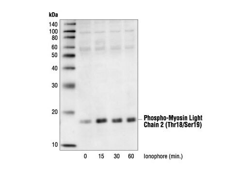
- Western blot analysis of extracts from HEK293 cells stimulated with calcium ionophore A23187 for the indicated times, using Phospho-MYL9 (Thr18/Ser19) Antibody (Product # PA5-17727).
Supportive validation
- Submitted by
- Invitrogen Antibodies (provider)
- Main image
- Experimental details
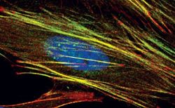
- Immunofluorescent analysis of Phospho-Myosin Light Chain 2 pThr18/Ser19 in HeLa cells using a Phospho-Myosin Light Chain 2 pThr18/Ser19 polyclonal antibody (Product # PA5-17727) (green). Actin filaments are labeled with a fluorescent red phalloidin. DNA is labeled using a fluorescent blue dye.

Supportive validation
- Submitted by
- Invitrogen Antibodies (provider)
- Main image
- Experimental details
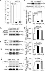
- Figure 1 Rnd3 is down-regulated in non-small lung cancer cell lines, H520 and H358. ( A ) Rnd3 mRNA detected by qRT-PCR is down-regulated in H358 and H520 compared to HBEC. ( B ) Rnd3 protein expression levels in cells by western blot. ( C ) Densitometry quantification of western band intensity in B. ( D ), ( F ), ( H ) & ( I ) A western blot to detect phosphorylated MYPT1, phosphorylated MLC2, ROCK1 and NICD in cells. ( E ), ( G ) & ( J ) Densitometry quantification of western band intensity showed up-regulation of Rho Kinase activity and NICD expression in H358 and H520 cells compared to HBEC. Western blots were quantified from three independent experimental repeats. BrdU-positive cells were quantified from 8 images taken from four slides. Data represent means +- S.D.

- Submitted by
- Invitrogen Antibodies (provider)
- Main image

- Experimental details
- Figure 3 Reintroduction of Rnd3 corrects Rho and Notch signaling in H520 and H358 cells. ( A ) & ( B ) Decreased NICD expression in H520-Rnd3 cells compared to H520 cells. ( C ) & ( D ) Decreased Rho Kinase activity in H520-Rnd3 cells compared to H520 cells detected by antibodies specific for pMYPT1 and pMLC2. ( E ) & ( F ) Decreased NICD expression in H358-Rnd3 cells compared to H358 cells. ( G ) & ( H ) Decreased Rho Kinase activity in H358-Rnd3 cells compared to H358 cells detected by antibodies specific for pMYPT1 and pMLC2. Western blots were quantified from three independent experimental repeats. Data represent means +- S.D.

- Submitted by
- Invitrogen Antibodies (provider)
- Main image

- Experimental details
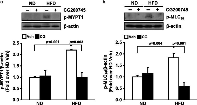
- Fig. 5 Effect of CG200745 on vascular contraction. Representative picture of western blot for phosphorylated MLC 20 at Thr18 and Ser19, phosphorylated MYPT1 at Thr853, and internal control beta-actin. Graphs summarize p-MYPT1 ( a ) and p-MLC 20 ( b ) in the mesenteric artery of ND and HFD-fed mice with/without CG200745 treatment. p-MYPT1 and p-MLC 20 were increased by HFD. CG200745 decreased phosphorylation of MYPT1 and MLC 20 . Data are presented as the mean +- SE ( n = 3-5 mice per group). p-, phosphorylated; MYPT1, Myosin phosphatase-targeting subunit1; MLC 20 , myosin light chain 20; ND, normal diet; HFD, high-fat diet; Veh, vehicle; CG, CG200745
- Submitted by
- Invitrogen Antibodies (provider)
- Main image

- Experimental details
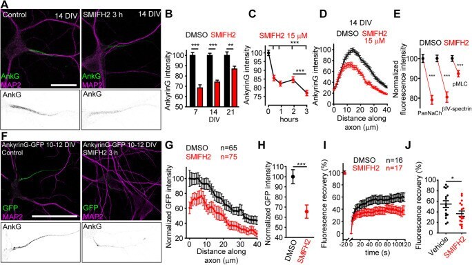
- Fig. 1 Formin inhibition decreases AIS protein density in hippocampal neurons. A A total of 14 DIV hippocampal neurons treated for 3 h with DMSO or 15 muM SMIFH2. AISs and somatodendritic compartment are identified by ankyrinG (green) and MAP2 (magenta). Bottom panels show AIS magnifications. Scale bar = 20 mum. B Normalized ankyrinG intensity in 7, 14, and 21 DIV hippocampal neurons treated for 3 h with vehicle (black bars) or 15 muM SMIFH2 (red bars). ** p < 0.01, *** p < 0.0001, Mann-Whitney test ( n = 150/experimental condition). C Normalized ankyrinG fluorescence intensity in 14 DIV neurons treated with DMSO (black dots) or 15 muM SMIFH2 (red dots) for 0.5, 1, 2, and 3 h. *** p < 0.001, Kruskal-Wallis, Dunn's multiple comparison test ( n = 150/experimental condition). D AnkyrinG intensity profile in 14 DIV neurons along the AIS. E Normalized AIS sodium channels (PanNaCh), betaIV-spectrin, and phospho-myosin light chain (pMLC) fluorescence intensity in 14 DIV neurons treated with DMSO (black) or 15 mM SMIFH2 (red) for 3 h. *** p < 0.001, unpaired t -test ( n = 120). F 10 DIV hippocampal neurons transfected with AnkyrinG-GFP for 48 h and exposed to DMSO or 15 muM SMIFH2 for 3 h. Bottom panels show AIS magnifications. Scale bar = 50 mum. G , H Normalized AIS GFP fluorescence intensity in vehicle (black) or SMIFH2 (red)-treated neurons ( H ) and GFP signal profile along the AIS ( G ). *** p < 0.001, unpaired t test. I Percentage of AIS AnkyrinG-GFP fluorescence recovery for