Explore
 Validate
 Learn
Learn Western blot
 Immunoprecipitation
Antibody data
- Antibody Data
- Antigen structure
- References [2]
- Comments [0]
- Validations
- Immunoprecipitation [1]
- Immunohistochemistry [1]
- Other assay [3]
Submit
Validation data
Reference
Comment
Report error
- Product number
- MA1-19287 - Provider product page

- Provider
- Invitrogen Antibodies
- Product name
- PAG1 Monoclonal Antibody (PAG-C1)
- Antibody type
- Monoclonal
- Antigen
- Synthetic peptide
- Description
- This antibody recognizes an epitope located in the intracellular C-terminal domain of Csk-binding protein (Cbp / PAG), a 46 kDa ubiquitously expressed transmembrane adaptor protein present in membrane rafts (glycosphingolipid-enriched microdomains), which however migrates on SDS PAGE gels anomalously as an 80 kDa molecule. A suggested positive control for Immunoprecipitation is RAJI human Burkitt lymphoma cell line. A suggested positive control for immunohistochemistry is appendix (germinal center of lymphatic follicle). For IHC staining, heat-mediated antigen retrieval in citrate buffer, pH 6.1, is recommended.
- Reactivity
- Human, Mouse, Rat, Bovine
- Host
- Mouse
- Isotype
- IgG
- Antibody clone number
- PAG-C1
- Vial size
- 100 μg
- Concentration
- 1 mg/mL
- Storage
- 4°C, do not freeze
Submitted references PAG1 directs SRC-family kinase intracellular localization to mediate receptor tyrosine kinase-induced differentiation.
Conformational coupling of integrin and Thy-1 regulates Fyn priming and fibroblast mechanotransduction.
Foltz L, Palacios-Moreno J, Mayfield M, Kinch S, Dillon J, Syrenne J, Levy T, Grimes M
Molecular biology of the cell 2020 Sep 15;31(20):2269-2282
Molecular biology of the cell 2020 Sep 15;31(20):2269-2282
Conformational coupling of integrin and Thy-1 regulates Fyn priming and fibroblast mechanotransduction.
Fiore VF, Strane PW, Bryksin AV, White ES, Hagood JS, Barker TH
The Journal of cell biology 2015 Oct 12;211(1):173-90
The Journal of cell biology 2015 Oct 12;211(1):173-90
No comments: Submit comment
Supportive validation
- Submitted by
- Invitrogen Antibodies (provider)
- Main image
- Experimental details
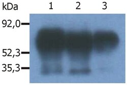
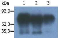
- Immunoprecipitation of human PAG/Cbp from the lysate of RAJI human Burkitt lymphoma cell line. Western blot was immunostained with anti-human PAG (MEM-255). Note: PAG/Cbp is a 46 kDa adaptor protein, which however migrates on SDS PAGE gels anomalously as an 80 kDa molecule. Lane 1,2: immunoprecipitation with anti-PAG (PAG-C1) Monoclonal antibody (Product # MA1-19287); Lane 3: immunoprecipitation with anti-PAG (polyclonal antibody).

Supportive validation
- Submitted by
- Invitrogen Antibodies (provider)
- Main image

- Experimental details
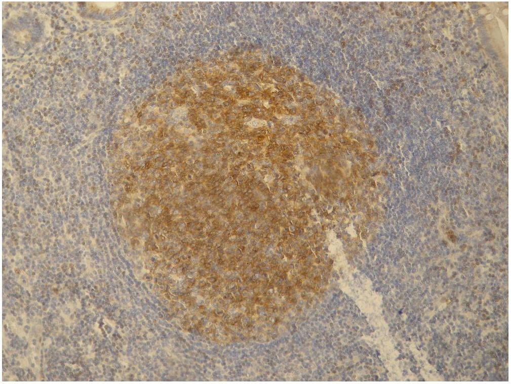
- Immunohistochemistry staining (paraffin sections) of PAG/Cbp in germinal center of lymphatic follicle and in dispersed T cells in human appendix tissue by Monoclonal antibody PAG-C1 (Product # MA1-19287). Positive signal in T cells.
Supportive validation
- Submitted by
- Invitrogen Antibodies (provider)
- Main image

- Experimental details
- Immunoprecipitation of PAG1 using a monoclonal antibody (Product # MA1-19287).
- Submitted by
- Invitrogen Antibodies (provider)
- Main image

- Experimental details
- Immunoprecipitation of human PAG/Cbp from the lysate of RAJI human Burkitt lymphoma cell line. Western blot was immunostained with anti-human PAG (MEM-255). Note: PAG/Cbp is a 46 kDa adaptor protein, which however migrates on SDS PAGE gels anomalously as an 80 kDa molecule. Lane 1,2: immunoprecipitation with anti-PAG (PAG-C1) Monoclonal antibody (Product # MA1-19287); Lane 3: immunoprecipitation with anti-PAG (polyclonal antibody).

- Submitted by
- Invitrogen Antibodies (provider)
- Main image
- Experimental details
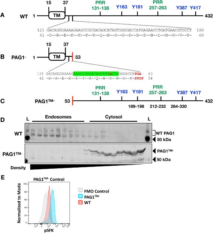
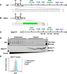
- FIGURE 1: SH-SY5Y cells expressing cytosolic PAG1 (PAG1 TM- ) were generated using CRISPR/Cas9. (A) WT PAG1 has a small extracellular N-terminal region, transmembrane domain (TM), and long intracellular C-terminal tail. Several phosphorylated residues and proline-rich regions (PRR, PXXP motif, green) act as docking sites for protein-protein interactions. Phosphorylated Y163/Y181 and Y387/417 (blue) are binding sites for FYN and LYN SH2 domains, respectively (; , ; ). (B) CRISPR sgRNA recognition site is highlighted in green. Sequencing results indicated a single base pair insertion that resulted in a stop codon to generate a heterozygous PAG1 - SH-SY5Y cell line (red). (C) Met residues upstream of the detected peptides act as alternate translation start sites to produce the cytosolic PAG1 product (PAG1 TM- ). PAG1 peptides were detected via mass spectrometry on PAG1 - cell lines (amino acid numbers are indicated in black). (D) Organelle fractionation comparing the endosomal distribution of WT PAG1 vs. PAG1 TM- . Fractions were taken from 25% to 2.5% iodixanol gradients and decrease in density from left to right. L, biotinylated ladder. PAG1 is extensively posttranslationally modified in WT cells and displays multiple bands on Western blots; in PAG1 TM- , a single band is visible, suggesting few such modifications. (E) Levels of activated SFKs were increased in PAG1 TM- -expressing SH-SY5Ys (blue) compared with WT SH-SY5Y cells (red). Cells were stained with a pSFK antibody re