Explore
 Validate
 Learn
Learn Western blot
 Immunohistochemistry
Antibody data
- Antibody Data
- Antigen structure
- References [2]
- Comments [0]
- Validations
- Western blot [1]
- Immunohistochemistry [1]
Submit
Validation data
Reference
Comment
Report error
- Product number
- HPA025224 - Provider product page

- Provider
- Atlas Antibodies
- Proper citation
- Atlas Antibodies Cat#HPA025224, RRID:AB_1856163
- Product name
- Anti-SDR16C5
- Antibody type
- Polyclonal
- Description
- Polyclonal Antibody against Human SDR16C5, Gene description: short chain dehydrogenase/reductase family 16C, member 5, Alternative Gene Names: RDH-E2, RDHE2, Validated applications: IHC, WB, Uniprot ID: Q8N3Y7, Storage: Store at +4°C for short term storage. Long time storage is recommended at -20°C.
- Reactivity
- Human
- Host
- Rabbit
- Conjugate
- Unconjugated
- Isotype
- IgG
- Vial size
- 100 µl
- Concentration
- 0.1 mg/ml
- Storage
- Store at +4°C for short term storage. Long time storage is recommended at -20°C.
- Handling
- The antibody solution should be gently mixed before use.
Submitted references LRG1 and SDR16C5 protein expressions differ according to HPV status in oropharyngeal squamous cell carcinoma.
Identification and Validation of Protein Biomarkers of Response to Neoadjuvant Platinum Chemotherapy in Muscle Invasive Urothelial Carcinoma
Randén-Brady R, Carpén T, Hautala LC, Tolvanen T, Haglund C, Joenväärä S, Mattila P, Mäkitie A, Lehtonen S, Hagström J, Silén S
Scientific reports 2024 Jun 19;14(1):14148
Scientific reports 2024 Jun 19;14(1):14148
Identification and Validation of Protein Biomarkers of Response to Neoadjuvant Platinum Chemotherapy in Muscle Invasive Urothelial Carcinoma
Vlahou A, Baras A, Gandhi N, Munari E, Faraj S, Shultz L, Marchionni L, Schoenberg M, Hahn N, Hoque M, Berman D, Bivalacqua T, Netto G
PLOS ONE 2015;10(7):e0131245
PLOS ONE 2015;10(7):e0131245
No comments: Submit comment
Enhanced validation
- Submitted by
- Atlas Antibodies (provider)
- Enhanced method
- Recombinant expression validation
- Main image
- Experimental details
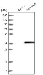
- Western blot analysis in control (vector only transfected HEK293T lysate) and SDR16C5 over-expression lysate (Co-expressed with a C-terminal myc-DDK tag (~3.1 kDa) in mammalian HEK293T cells, LY408444).
- Sample type
- Human
- Protocol
- Protocol

Supportive validation
- Submitted by
- Atlas Antibodies (provider)
- Enhanced method
- Orthogonal validation
- Main image
- Experimental details
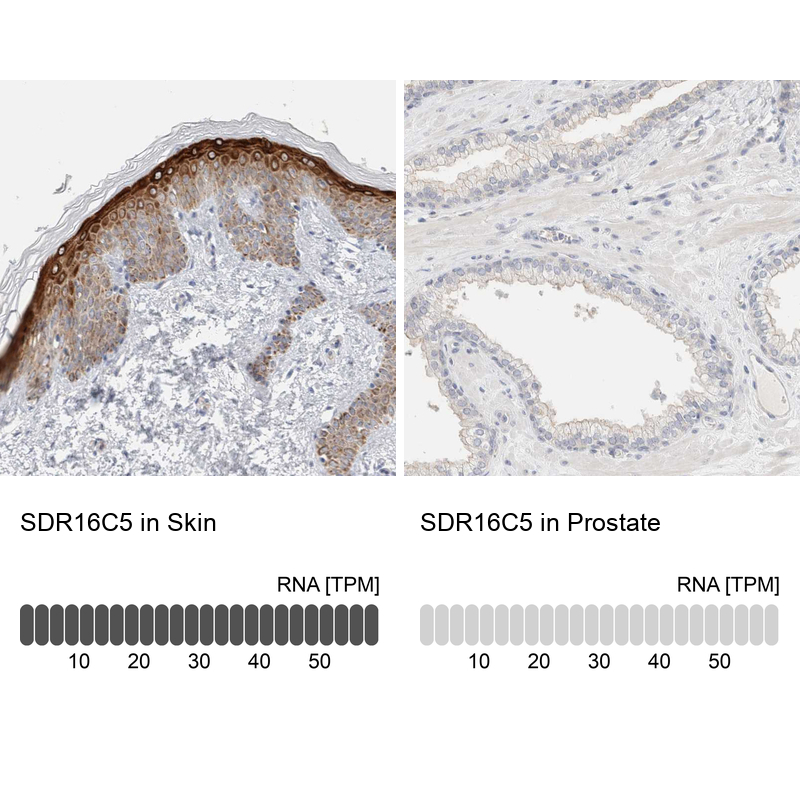
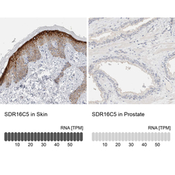
- Immunohistochemistry analysis in human skin and prostate tissues using HPA025224 antibody. Corresponding SDR16C5 RNA-seq data are presented for the same tissues.
- Sample type
- Human
- Protocol
- Protocol