Explore
 Validate
 Learn
Learn Western blot
 ELISA
ELISAAntibody data
- Antibody Data
- Antigen structure
- References [1]
- Comments [0]
- Validations
- Western blot [1]
- Immunocytochemistry [2]
- Immunohistochemistry [2]
Submit
Validation data
Reference
Comment
Report error
- Product number
- MA1-19344 - Provider product page

- Provider
- Invitrogen Antibodies
- Product name
- Blood Group Lewis A Monoclonal Antibody (7LE)
- Antibody type
- Monoclonal
- Antigen
- Other
- Description
- This antibody recognizes Lewisa blood group antigen, a carbohydrate determinant carried on both glycolipids and glycoproteins, expressed on colonic epithelial cells. Lewisa may be useful for detection of gastrointestinal, pancreatic and colorectal tumors. For IHC (P), heat mediated antigen retrieval in sodium citrate is recommended.
- Reactivity
- Human
- Host
- Mouse
- Isotype
- IgG
- Antibody clone number
- 7LE
- Vial size
- 100 μg
- Concentration
- 1 mg/mL
- Storage
- 4°C, do not freeze
Submitted references High-throughput carbohydrate microarray profiling of 27 antibodies demonstrates widespread specificity problems.
Manimala JC, Roach TA, Li Z, Gildersleeve JC
Glycobiology 2007 Aug;17(8):17C-23C
Glycobiology 2007 Aug;17(8):17C-23C
No comments: Submit comment
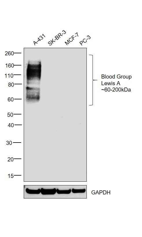
Supportive validation
- Submitted by
- Invitrogen Antibodies (provider)
- Main image
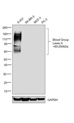
- Experimental details
- Western blot was performed using Anti-Blood Group Lewis A Monoclonal Antibody (Product # MA1-19344) and bands spanning between 60-200 kDa corresponding to glycosylated forms of Blood Group Lewis A were observed in A-431 and were absent in SK-BR-3, MCF-7 and PC-3 cell lysates. Whole cell extracts (30 µg lysate) of A-431 (Lane 1), SK-BR-3 (Lane 2), MCF-7 (Lane 3) and PC-3 (Lane 4) were electrophoresed using Novex® NuPAGE® 4-12 % Bis-Tris gel (Product # NP0322BOX). Resolved proteins were then transferred onto a nitrocellulose membrane (Product # IB23001) by iBlot® 2 Dry Blotting System (Product # IB21001). The blot was probed with the primary antibody (1:1000 dilution) and detected by chemiluminescence with Goat anti-Mouse IgG (H+L) Superclonal™ Recombinant Secondary Antibody, HRP (Product # A28177, 1:4000 dilution) using the iBright FL 1000 (Product # A32752). Chemiluminescent detection was performed using Novex® ECL Chemiluminescent Substrate Reagent Kit (Product # WP20005).
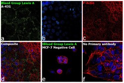
Supportive validation
- Submitted by
- Invitrogen Antibodies (provider)
- Main image

- Experimental details
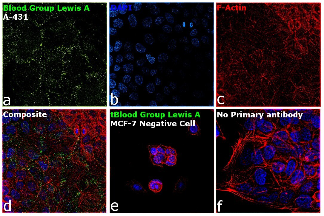
- Immunofluorescence analysis of Blood Group Lewis A was performed using 70% confluent log phase A-431 cells. The cells were fixed with 4% paraformaldehyde for 10 minutes, permeabilized with 0.1% Triton™ X-100 for 15 minutes, and blocked with 1% BSA for 1 hour at room temperature. The cells were labeled with Blood Group Lewis A Monoclonal Antibody (7LE) (Product # MA1-19344) at 1:100 dilution in 0.1% BSA, incubated at 4 degree Celsius overnight and then labeled with Goat anti-Mouse IgG (H+L), Superclonal™ Recombinant Secondary Antibody, Alexa Fluor 488 conjugate (Product # A28175) at a dilution of 1:2000 for 45 minutes at room temperature (Panel a: green). Nuclei (Panel b: blue) were stained with SlowFade® Gold Antifade Mountant with DAPI (Product # S36938). F-actin (Panel c: red) was stained with Rhodamine Phalloidin (Product # R415, 1:300). Panel d represents the merged image showing plasma membrane and cytoplasmic localization. Panel e shows MCF-7 cells with no expression of Blood Group Lewis A. Panel f represents control cells with no primary antibody to assess background. The images were captured at 60X magnification.

- Submitted by
- Invitrogen Antibodies (provider)
- Main image
- Experimental details
- Immunofluorescence analysis of Blood Group Lewis A was performed using 70% confluent log phase A-431 cells. The cells were fixed with 4% paraformaldehyde for 10 minutes, permeabilized with 0.1% Triton™ X-100 for 15 minutes, and blocked with 1% BSA for 1 hour at room temperature. The cells were labeled with Blood Group Lewis A Monoclonal Antibody (7LE) (Product # MA1-19344) at 1:100 dilution in 0.1% BSA, incubated at 4 degree Celsius overnight and then labeled with Goat anti-Mouse IgG (H+L), Superclonal™ Recombinant Secondary Antibody, Alexa Fluor 488 conjugate (Product # A28175) at a dilution of 1:2000 for 45 minutes at room temperature (Panel a: green). Nuclei (Panel b: blue) were stained with SlowFade® Gold Antifade Mountant with DAPI (Product # S36938). F-actin (Panel c: red) was stained with Rhodamine Phalloidin (Product # R415, 1:300). Panel d represents the merged image showing plasma membrane and cytoplasmic localization. Panel e shows MCF-7 cells with no expression of Blood Group Lewis A. Panel f represents control cells with no primary antibody to assess background. The images were captured at 60X magnification.
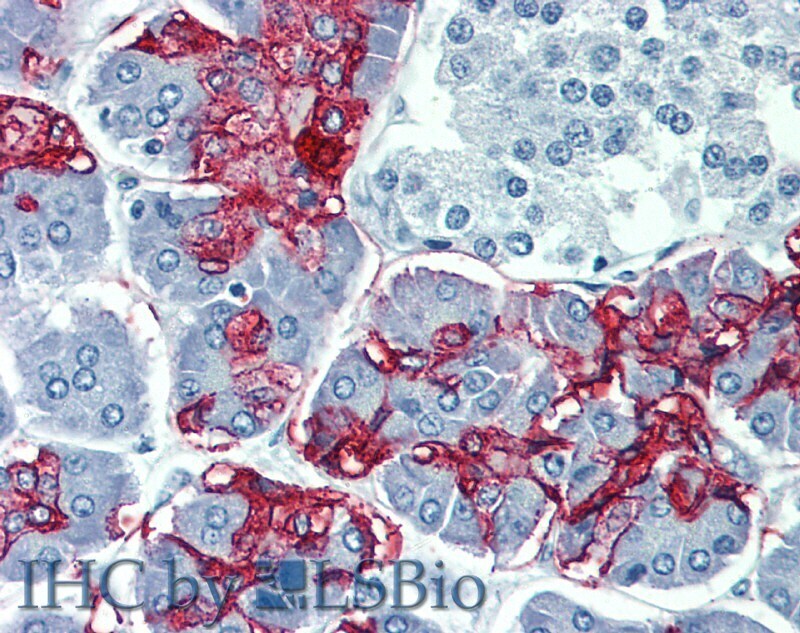
Supportive validation
- Submitted by
- Invitrogen Antibodies (provider)
- Main image

- Experimental details
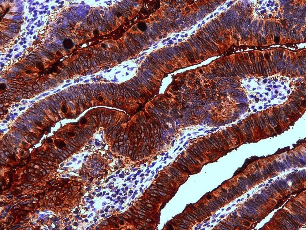
- Immunohistochemistry staining of human colon adenocarcinoma (paraffin-embedded sections) with anti-Blood Group Lewis a (7LE) Monoclonal antibody (Product # MA1-19344).
- Submitted by
- Invitrogen Antibodies (provider)
- Main image
- Experimental details
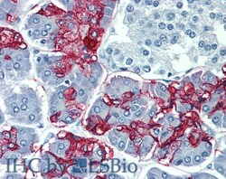
- Immunohistochemistry staining of human pancreas (paraffin-embedded sections) with anti-Blood Group Lewis a (7LE) Monoclonal antibody (Product # MA1-19344).