 Explore
 Validate
 Learn
Learn Western blot
Antibody data
- Antibody Data
- Antigen structure
- References [0]
- Comments [0]
- Validations
- Western blot [7]
- Immunocytochemistry [1]
Submit
Validation data
Reference
Comment
Report error
- Product number
- PA5-78365 - Provider product page

- Provider
- Invitrogen Antibodies
- Product name
- GABARAP Polyclonal Antibody
- Antibody type
- Polyclonal
- Antigen
- Synthetic peptide
- Description
- Positive Control: 293T, Jurkat, Raji, NCI-H929, A431, HeLa, human GABARAP-transfected 293T Predicted Reactivity: Mouse (100%), Rat (100%), Pig (100%), Rabbit (100%), Rhesus Monkey (100%), Bovine (100%) Store product as a concentrated solution. Centrifuge briefly prior to opening the vial.
- Reactivity
- Human
- Host
- Rabbit
- Isotype
- IgG
- Vial size
- 100 µL
- Concentration
- 1 mg/mL
- Storage
- Store at 4°C short term. For long term storage, store at -20°C, avoiding freeze/thaw cycles.
No comments: Submit comment
Supportive validation
- Submitted by
- Invitrogen Antibodies (provider)
- Main image

- Experimental details
- Western blot analysis of GABARAP in A) 293T whole cell lysate, B) whole cell lysate of human GABARAP-transfected 293T cells using 30 µg of protein. Samples were separated with 15% SDS-PAGE and incubated with GABARAP polyclonal antibody (Product # PA5-78365) using a dilution of 1:10,000.

- Submitted by
- Invitrogen Antibodies (provider)
- Main image

- Experimental details
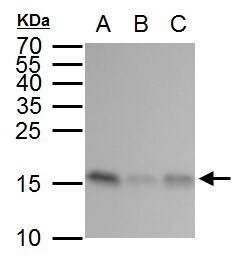
- Western blot analysis of GABARAP in A) 293T whole cell lysate, B) A431 whole cell lysate, C) HeLa whole cell lysate using 30 µg of protein. Samples were separated with 15% SDS-PAGE and incubated with GABARAP polyclonal antibody (Product # PA5-78365) using a dilution of 1:2000.

- Submitted by
- Invitrogen Antibodies (provider)
- Main image

- Experimental details
- Western blot analysis of GABARAP in non-transfected (-) and transfected (+) 293T cells using 30 µg of protein. Samples were separated with 15% SDS-PAGE and incubated with GABARAP polyclonal antibody (Product # PA5-78365) using a dilution of 1:1000.

- Submitted by
- Invitrogen Antibodies (provider)
- Main image

- Experimental details
- GABARAP Polyclonal Antibody detects GABARAP protein by western blot analysis. A. 30 µg 293T whole cell lysate/extract. B. 30 µg A431 whole cell lysate/extract. C. 30 µg HeLa whole cell lysate/extract.15 % SDS-PAGE. GABARAP Polyclonal Antibody (Product # PA5-78365) dilution: 1:2,000.
- Submitted by
- Invitrogen Antibodies (provider)
- Main image

- Experimental details
- GABARAP Polyclonal Antibody detects GABARAP protein by western blot analysis. A. 30 µg 293T whole cell lysate/extract. B. 30 whole cell lysate/extract of human GABARAP -transfected 293T cells.15 % SDS-PAGE. GABARAP Polyclonal Antibody (Product # PA5-78365) dilution: 1:10,000.

- Submitted by
- Invitrogen Antibodies (provider)
- Main image

- Experimental details
- GABARAP Polyclonal Antibody detects GABARAP protein by western blot analysis. A. 30 µg Jurkat whole cell lysate/extract. B. 30 µg Raji whole cell lysate/extract. C. 30 µg NCI-H929 whole cell lysate/extract.15 % SDS-PAGE. GABARAP Polyclonal Antibody (Product # PA5-78365) dilution: 1:2,000.

- Submitted by
- Invitrogen Antibodies (provider)
- Main image

- Experimental details
- Western blot was performed using Anti-Gamma-aminobutyric acid receptor-associated protein Polyclonal Antibody (Product # PA5-78365) and 13.9 kDa band corresponding to Gamma-aminobutyric acid receptor-associated protein was observed along with an uncharacterized band (*) at ~80 kDa. Chloroquine is known to increase the expression of Gamma-aminobutyric acid receptor-associated protein. Membrane enriched cell extracts (30 µg lysate) of A549 (Lane 1) and A549 treated with chloroquine (50uM/ml for 20 hr) (Lane 2), NIH/3T3 (Lane 3) and NIH/3T3 treated with chloroquine (50uM/ml for 20 hr) (Lane 4), HeLa (Lane 5) and HeLa treated with chloroquine (50uM/ml for 20 hr) (Lane 6), LNCaP (Lane 7), Mouse brain (Lane 8) and Mouse skeletal muscle (Lane 9) were electrophoresed using Novex® NuPAGE® 4-12% Bis-Tris gel (Product # NP0342BOX). Resolved proteins were then transferred onto a nitrocellulose membrane (Product # IB23001) by iBlot® 2 Dry Blotting System (Product # IB21001). The blot was probed with the primary antibody (1:5000 dilution) and detected by Goat anti-Rabbit IgG (H+L) Superclonal™ Recombinant Secondary Antibody, HRP conjugate (Product # A27036, 1:4000 dilution) using the iBright FL 1000 (Product # A32752). Chemiluminescent detection was performed using Novex® ECL Chemiluminescent Substrate Reagent Kit (Product # WP20005).

Supportive validation
- Submitted by
- Invitrogen Antibodies (provider)
- Main image

- Experimental details
- GABARAP Polyclonal Antibody detects GABARAP protein at cytoplasm by immunofluorescent analysis. Sample: U2OS cells were fixed in ice-cold MeOH for 5 min. Green: GABARAP protein stained by GABARAP Polyclonal Antibody (Product # PA5-78365) diluted at 1:500. Blue: Hoechst 33342 staining.
