Explore
 Validate
 Learn
Learn Western blot
 Immunocytochemistry
Antibody data
- Antibody Data
- Antigen structure
- References [3]
- Comments [0]
- Validations
- Immunocytochemistry [2]
- Flow cytometry [1]
- Other assay [2]
Submit
Validation data
Reference
Comment
Report error
- Product number
- 720161 - Provider product page

- Provider
- Invitrogen Antibodies
- Product name
- Annexin A6 Polyclonal Antibody
- Antibody type
- Polyclonal
- Antigen
- Recombinant full-length protein
- Description
- These Polyclonal antibodies are of rabbit origin developed by immunizing animals with proteins or peptides. The polyclonal antibody is purified by affinity purification from the rabbit sera generated after immunizing the rabbits with a specific type of protein or peptide. The purified antibody is tested for its functionality in various relevant research applications. The antibody is developed for Research Use Only and is non-hazardous or non-infectious in nature. This antibody is predicted to react with Monkey, Rat, Mouse and Rabbit.
- Reactivity
- Human, Mouse
- Host
- Rabbit
- Isotype
- IgG
- Vial size
- 100 μg
- Concentration
- 0.5 mg/mL
- Storage
- Store at 4°C short term. For long term storage, store at -20°C, avoiding freeze/thaw cycles.
Submitted references Enterohemorrhagic Escherichia coli Effector Protein EspF Interacts With Host Protein ANXA6 and Triggers Myosin Light Chain Kinase (MLCK)-Dependent Tight Junction Dysregulation.
Treatment with galectin-1 improves myogenic potential and membrane repair in dysferlin-deficient models.
Chemotherapy elicits pro-metastatic extracellular vesicles in breast cancer models.
Hua Y, Wu J, Fu M, Liu J, Li X, Zhang B, Zhao W, Wan C
Frontiers in cell and developmental biology 2020;8:613061
Frontiers in cell and developmental biology 2020;8:613061
Treatment with galectin-1 improves myogenic potential and membrane repair in dysferlin-deficient models.
Vallecillo-Zúniga ML, Rathgeber MF, Poulson PD, Hayes S, Luddington JS, Gill HN, Teynor M, Kartchner BC, Valdoz J, Stowell C, Markham AR, Arthur C, Stowell S, Van Ry PM
PloS one 2020;15(9):e0238441
PloS one 2020;15(9):e0238441
Chemotherapy elicits pro-metastatic extracellular vesicles in breast cancer models.
Keklikoglou I, Cianciaruso C, Güç E, Squadrito ML, Spring LM, Tazzyman S, Lambein L, Poissonnier A, Ferraro GB, Baer C, Cassará A, Guichard A, Iruela-Arispe ML, Lewis CE, Coussens LM, Bardia A, Jain RK, Pollard JW, De Palma M
Nature cell biology 2019 Feb;21(2):190-202
Nature cell biology 2019 Feb;21(2):190-202
No comments: Submit comment
Supportive validation
- Submitted by
- Invitrogen Antibodies (provider)
- Main image

- Experimental details
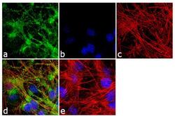
- Immunofluorescence was performed on fixed and permeabilized C2C12 cells for detection of Annexin A6 using Anti-Annexin A6 Rabbit Polyclonal Antibody (Product # 720161, 1 µg/mL) and labeled with Goat anti-Rabbit IgG (H+L) Superclonal™ Secondary Antibody, Alexa Fluor® 488 conjugate (Product # A27034, 1:2000). Panel a) shows representative cells that were stained for detection and localization of Annexin A6 protein (green), Panel b) is stained for nuclei (blue) using SlowFade® Gold Antifade Mountant with DAPI (Product # S36938). Panel c) represents cytoskeletal F-actin staining using Alexa Fluor® 555 Rhodamine Phalloidin (Product # R415, 1:300). Panel d) is a composite image of Panels a, b and c clearly demonstrating cytoplasmic localization of Annexin A6 Panel e) represents control cells with no primary Antibody to assess background.

- Submitted by
- Invitrogen Antibodies (provider)
- Main image
- Experimental details
- Immunofluorescence was performed on fixed and permeabilized C2C12 cells for detection of Annexin A6 using Anti-Annexin A6 Rabbit Polyclonal Antibody (Product # 720161, 1 µg/mL) and labeled with Goat anti-Rabbit IgG (Heavy Chain) Superclonal™ Secondary Antibody, Alexa Fluor® 488 conjugate (Product # A27034, 1:2000). Panel a) shows representative cells that were stained for detection and localization of Annexin A6 protein (green), Panel b) is stained for nuclei (blue) using SlowFade® Gold Antifade Mountant with DAPI (Product # S36938). Panel c) represents cytoskeletal F-actin staining using Alexa Fluor® 555 Rhodamine Phalloidin (Product # R415, 1:300). Panel d) is a composite image of Panels a, b and c clearly demonstrating cytoplasmic localization of Annexin A6 Panel e) represents control cells with no primary Antibody to assess background.

Supportive validation
- Submitted by
- Invitrogen Antibodies (provider)
- Main image
- Experimental details
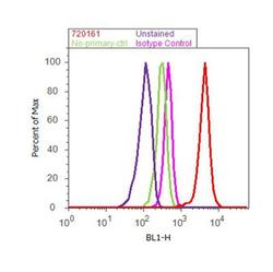
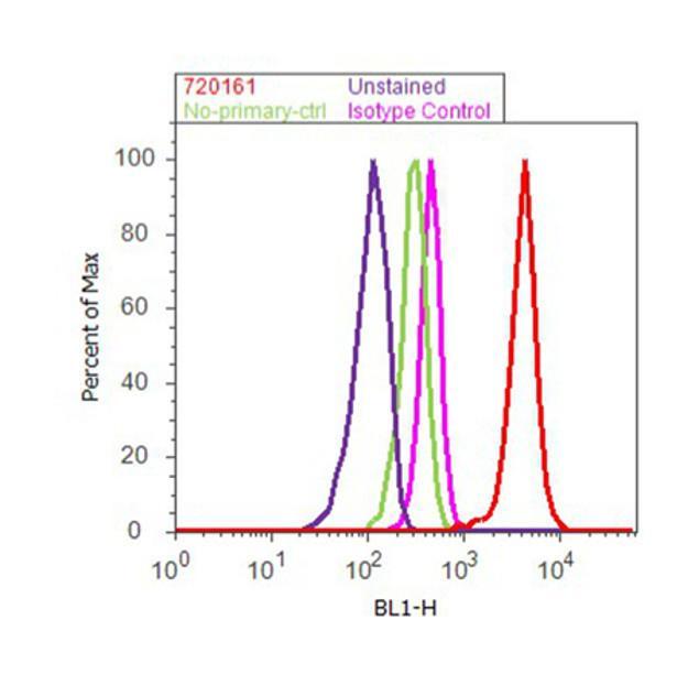
- Flow Cytometry analysis of Annexin A6 was performed on K562 cells labeled with Anti-Annexin A6 Rabbit Polyclonal Antibody (Product# 720161, 2-4 ug/ 1M cells) or with Rabbit isotype control and detected with Goat anti-Rabbit IgG (H+L) Superclonal™ Secondary Antibody, (Alexa Fluor® 488 conjugate, Product # A27034, 0.4 ug/ml, 1:2500) as represented by the red and pink histograms respectively. The purple histogram represents unstained control cells and the green histogram represents no-primary-Antibody control. A representative of 10,000 cells were acquired and analyzed for each sample using an Attune® Acoustic Focusing Cytometer (4468770).
Supportive validation
- Submitted by
- Invitrogen Antibodies (provider)
- Main image
- Experimental details
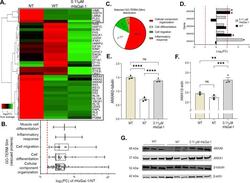
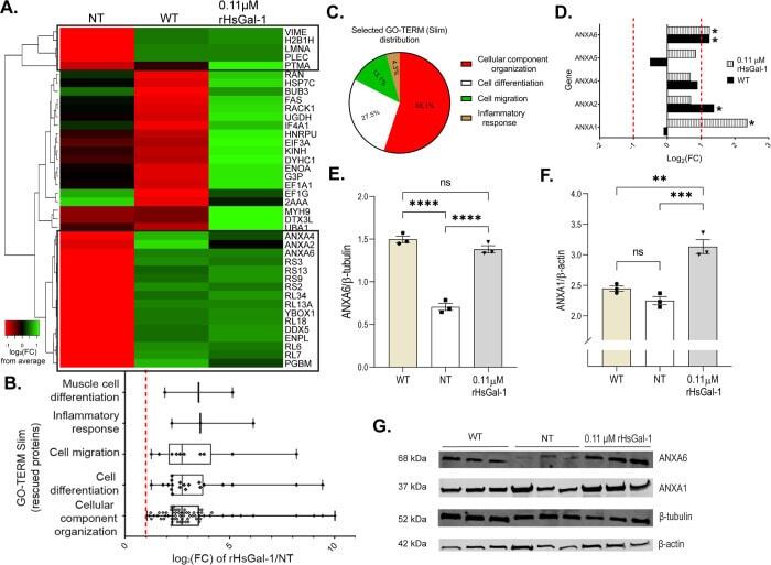
- Fig 6 Shotgun proteomic analyses (MS/MS) of AJ WT, AJ -/- NT and 0.11 muM rHsGal-1 treated myotubes. A. Heatmap of top 20 and bottom 20 expressed proteins for WT myotubes based on Log 2 fold change (FC) from the mean value all conditions. Box denotes cluster of proteins that had similar FC values between WT and rHsGal-1 treatment. B. GO-Term Slim analysis of rescued proteins. C. Relative amount of proteins found in the selected GO-Terms(B). D. Log 2 FC of Annexin family proteins (calculated based on WT and Gal-1 FC from NT). E. Quantification of ANXA6 after 48h differentiation in WT, NT, and 0.11 muM rHsGal-1 treated myotubes. F. Quantification of ANXA1 after 48h in WT, NT, and 0.11 muM rHsGal-1 treated myotubes. G. Western blot images of ANXA1 and ANXA6. p values were measured by 2-way ANOVA multiple comparison test and indicated by *p< 0.05, **p< 0.01, and ***p< 0.001. Error bars represent SEM. n = 3. All proteins were preselected based on protein ID significance values of >=20% (p
- Submitted by
- Invitrogen Antibodies (provider)
- Main image
- Experimental details
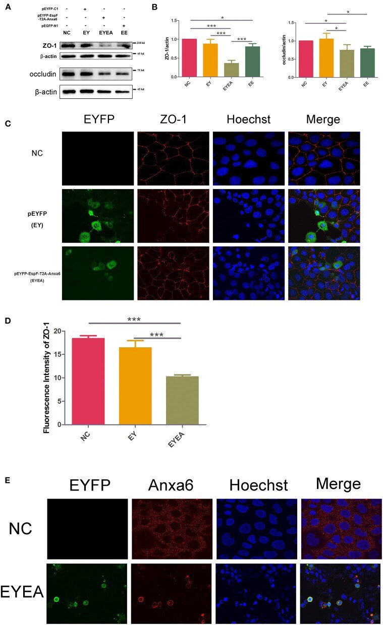
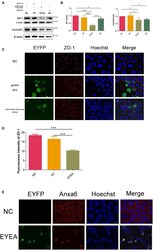
- Figure 5 The co-expression of EspF and ANXA6 protein in Caco-2 cells reduced the level of ZO-1 and occludin and disrupted the structure of ZO-1. (A,B) The expression levels and grayscale analysis of WB strips in Caco-2 cells transfected with plasmids as indicated. The grayscale analysis was performed in ImageJ software. Error bars represent means +- SD from three independent experiments. (C,D) Immunofluorescence and quantification analysis of ZO-1 protein. Nucleus was labeled with Hoechst in blue. ZO-1 was labeled with anti-ZO-1 antibody in red. Green indicates fluorescence expressed by the fluorescent plasmid. Images were acquired on an FV1000 confocal laser scanning microscope using a 60X oil objective. The fluorescence intensity of ZO-1 was analyzed by ImageJ software. Error bars represent means +- SD from three independent experiments. *Statistically significant difference. * p < 0.05. *** p < 0.001. (E) Immunofluorescence of ANXA6 protein in Caco-2 cells. As described in (C,D) ANXA6 was labeled with an anti-ANXA6 antibody in red. Images were acquired on an FV1000 confocal laser scanning microscope using a 60X oil objective. NC, non-transfected cells; EYEA, cells transfected with pEYFP-EspF-T2A-ANXA6.