Explore
 Validate
 Learn
Learn Western blot
 Flow cytometry
Antibody data
- Antibody Data
- Antigen structure
- References [1]
- Comments [0]
- Validations
- Western blot [4]
- Immunocytochemistry [1]
- Other assay [3]
Submit
Validation data
Reference
Comment
Report error
- Product number
- MA5-25133 - Provider product page

- Provider
- Invitrogen Antibodies
- Product name
- BMAL1 Monoclonal Antibody (OTI3G9)
- Antibody type
- Monoclonal
- Antigen
- Recombinant full-length protein
- Reactivity
- Human, Mouse, Rat, Canine
- Host
- Mouse
- Isotype
- IgG
- Antibody clone number
- OTI3G9
- Vial size
- 100 µL
- Concentration
- 1 mg/mL
- Storage
- -20° C, Avoid Freeze/Thaw Cycles
Submitted references BMAL1 attenuates intracerebral hemorrhage-induced secondary brain injury in rats by regulating the Nrf2 signaling pathway.
Gong Y, Zhang G, Li B, Cao C, Cao D, Li X, Li H, Ye M, Shen H, Chen G
Annals of translational medicine 2021 Nov;9(21):1617
Annals of translational medicine 2021 Nov;9(21):1617
No comments: Submit comment
Supportive validation
- Submitted by
- Invitrogen Antibodies (provider)
- Main image

- Experimental details
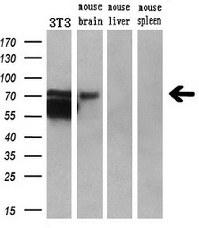
- Western blot analysis of ARNTL in 3T3 cells, mouse brain, mouse liver, mouse spleen lysate using 10 µg per lane. Samples were probed with ARNTL (Product # MA5-25133) monoclonal antibody at a dilution of 1:200.
- Submitted by
- Invitrogen Antibodies (provider)
- Main image
- Experimental details
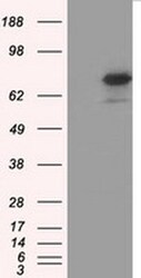
- Western blot analysis of ARNTL in HEK293T cells in untransfected (Left lane) and transfected (Right lane) samples using 5 µg per lane. The samples were separated by SDS-PAGE and probed with ARNTL (Product # MA5-25133) monoclonal antibody.
- Submitted by
- Invitrogen Antibodies (provider)
- Main image
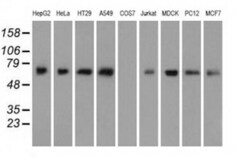
- Experimental details
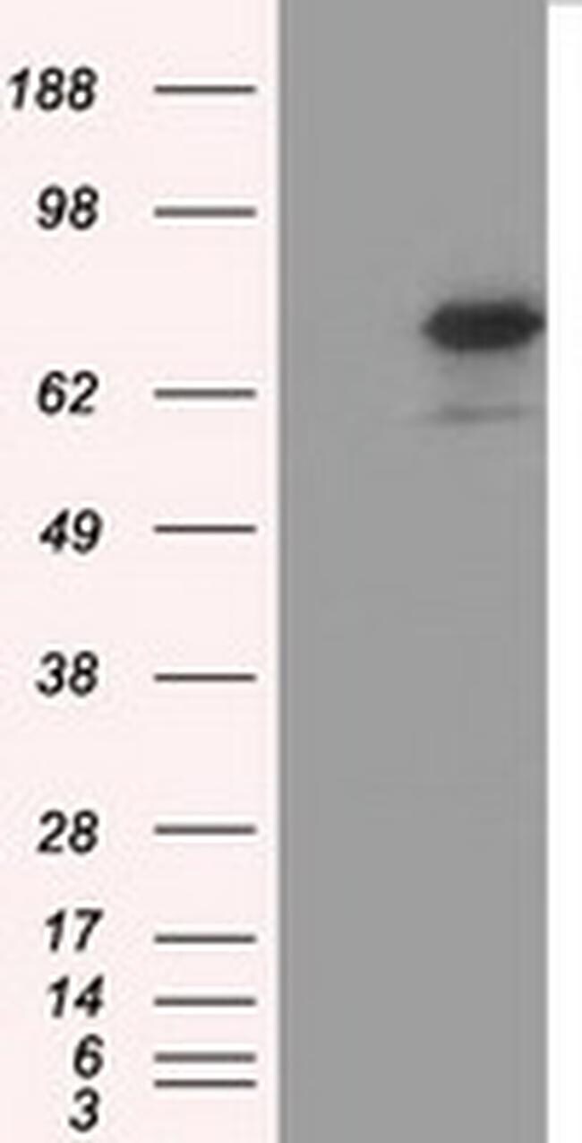
- Western blot analysis of ARNTL in HepG2, HeLa, HT29, A549, COS7, Jurkat, MDCK, PC12, MCF7 cells using 35 µg per lane. Samples were probed with ARNTL (Product # MA5-25133) monoclonal antibody.
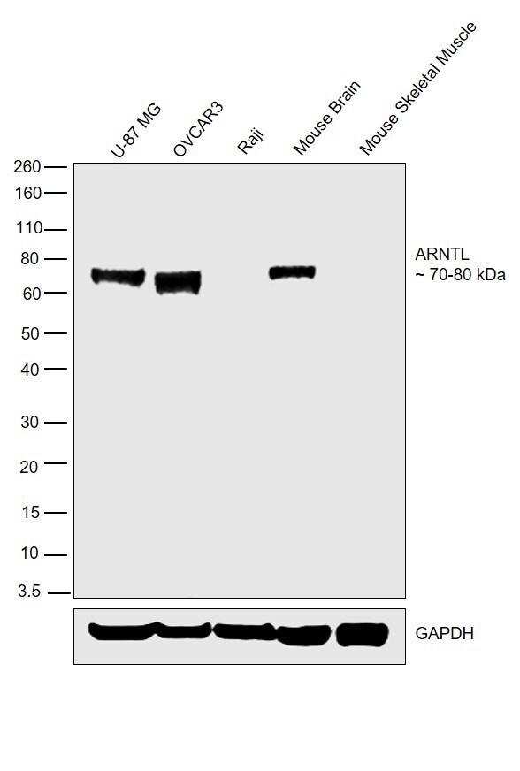
- Submitted by
- Invitrogen Antibodies (provider)
- Main image
- Experimental details
- Western blot was performed using Anti-BMAL1 Monoclonal Antibody (OTI3G9) (Product # MA5-25133) and bands around 70-80 kDa corresponding to BMAL1/ARNTL were observed in U-87 MG, OVCAR3 and Mouse Brain but not in Raji or Mouse Skeletal Muscle. Whole cell extracts (40 µg lysate) of U-87 MG (Lane 1), OVCAR3 (Lane 2), Raji (Lane 3), Mouse Brain (Lane 4) or Mouse Skeletal Muscle (Lane 5) were electrophoresed using NuPAGE® 4-12 % Bis-Tris gel (Product # NP0322BOX). Resolved proteins were then transferred onto a nitrocellulose membrane (Product # IB23001) by iBlot® 2 Dry Blotting System (Product # IB21001). The blot was probed with the primary antibody (1:1000 dilution) and detected by chemiluminescence with Goat anti-Mouse IgG (H+L), Superclonal™ Recombinant Secondary Antibody, HRP (Product # A28177, 1:4000 dilution) using the iBright FL 1000 (Product # A32752). Chemiluminescent detection was performed using Novex® ECL Chemiluminescent Substrate Reagent Kit (Product # WP20005).
Supportive validation
- Submitted by
- Invitrogen Antibodies (provider)
- Main image

- Experimental details
- Immunofluorescent analysis of ARNTL in COS7 cells. Cells were transfected with a plasmid overexpressing ARNTL and probed with a ARNTL monoclonal antibody (Product # MA5-25133).

Supportive validation
- Submitted by
- Invitrogen Antibodies (provider)
- Main image
- Experimental details
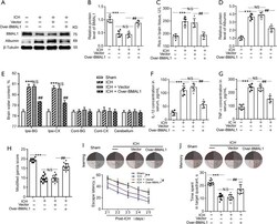
- Effects of BMAL1 overexpression on oxidative stress, BBB injury, brain edema, inflammation and neurobehaviors after ICH. (A) Western blot analysis of levels of the BMAL1 and Albumin proteins (index of BBB injury) in the sham, ICH, ICH + Vector and ICH + Over-BMAL1 groups at 24 h after ICH. (B) Quantification of the relative BMAL1 protein levels in the four groups. (C) ROS levels in the brain tissues from rats in the four groups. (D) Quantification of the relative Albumin protein levels in the four groups. (E) The effects of BMAL1 overexpression on the brain water content of rats in the four groups. (F) Concentrations of IL-1beta in the serum of the sham ICH, ICH + Vector and ICH+Over-BMAL1 groups at 24 h after ICH. (G) Concentrations of TNF-alpha in the serum of the four groups listed above. (H) The scores on the modified Garcia test in the sham, ICH, ICH + Vector and ICH + Over-BMAL1 groups at 24 h after ICH. (I) Representative images illustrate swimming trajectories at 25 d (learning) and escape latency in the Morris water maze test at 21 to 25 d after ICH in the sham, ICH, ICH + Vector and ICH + Over BMAL1 rats. (J) Representative images illustrate swimming trajectories at 26 d (memory) and time spent in the platform quadrant in the Morris water maze test at 26 d after ICH in the groups listed above. **P

- Submitted by
- Invitrogen Antibodies (provider)
- Main image
- Experimental details
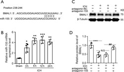
- The miR-155 expression level and level of the BMAL1 protein by inhibiting miR-155 after ICH. (A) The binding site of BMAL1 in the miR-155 3'UTR. (B) The levels of miR-155 were increased after ICH, and the increase was established at 3 h after ICH and remained at a high level. (C) Western blot analysis of the BMAL1 protein levels in the sham, ICH, ICH + antagomir-NC and ICH + antagomir-155 groups. (D) Quantification of the relative BMAL1 protein levels in the groups listed above. All data are presented as the means +- SEM, *P

- Submitted by
- Invitrogen Antibodies (provider)
- Main image

- Experimental details
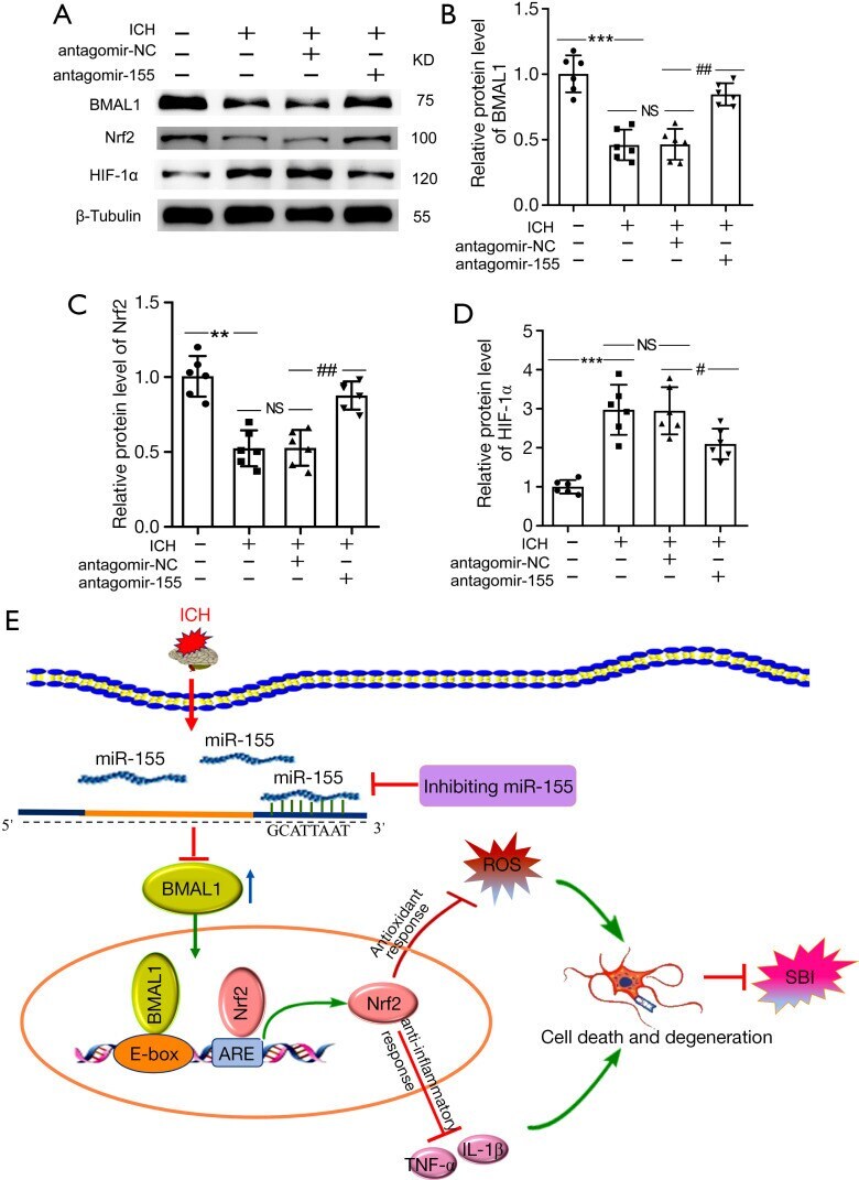
- Effects of increased BMAL1 on the Nrf2 signaling pathway after ICH. (A) Western blot analysis of the relative levels of the BMAL1, Nrf2 and HIF-1alpha proteins in the sham, ICH, ICH + antagomir-NC and ICH + antagomir-155 groups at 24 h after ICH; (B) Western blot analysis of the relative levels of the BMAL1 protein in the groups listed above; (C) Western blot analysis of the relative levels of the Nrf2 protein in the groups listed above; (D) Western blot analysis of the relative levels of the HIF-1alpha protein in the groups listed above; (E) Schematic representation of the role and related mechanism of BMAL1 in SBI after ICH. The BMAL1 protein level decreased in the brain tissue of rats after ICH. After antagomir-155 treatment, the BMAL1 protein was upregulated, and then the Nrf2 signaling pathway was activated to attenuate SBI induced by ICH, including oxidative stress, inflammation, and neuronal death. All data are presented as the mean +- SEM. ***P