 Explore
 Validate
 Learn
Learn Western blot
Antibody data
- Antibody Data
- Antigen structure
- References [6]
- Comments [0]
- Validations
- Western blot [2]
- Immunocytochemistry [2]
- Flow cytometry [1]
- Other assay [2]
Submit
Validation data
Reference
Comment
Report error
- Product number
- PA1-523 - Provider product page

- Provider
- Invitrogen Antibodies
- Product name
- BMAL1 Polyclonal Antibody
- Antibody type
- Polyclonal
- Antigen
- Synthetic peptide
- Description
- PA1-523 detects BMAL1/aryl hydrocarbon nuclear translocator 3 (ARNT3) from hamster and mouse tissues as well as recombinant human BMAL1. PA1-523 has been sucessfully used in Western blot procedures. By Western blot, this antibody detects a 110 kDa protein which corresponds to the product of a hamster GST-BMAL1 fusion construct overexpressed in E. coli. This antibody detects a non-specific band in U87-MG cell lysates at ~105kDa and in NIH-3T3 cell lysates at ~135kDa. PA1-523 immunizing peptide corresponds to amino acid residues 582-594 from mouse BMAL1. This sequence is completely conserved between mouse, rat, guinea pig, and human BMAL1. PA1-523 immunizing peptide (Cat. # PEP-075) is available for use in neutralization and control experiments.
- Reactivity
- Human, Mouse, Rat, Hamster
- Host
- Rabbit
- Isotype
- IgG
- Vial size
- 200 μL
- Concentration
- 1 mg/mL
- Storage
- -20°C, Avoid Freeze/Thaw Cycles
Submitted references Prolonged Administration of Melatonin Ameliorates Liver Phenotypes in Cholestatic Murine Model.
Melatonin modulates daytime-dependent synaptic plasticity and learning efficiency.
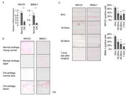
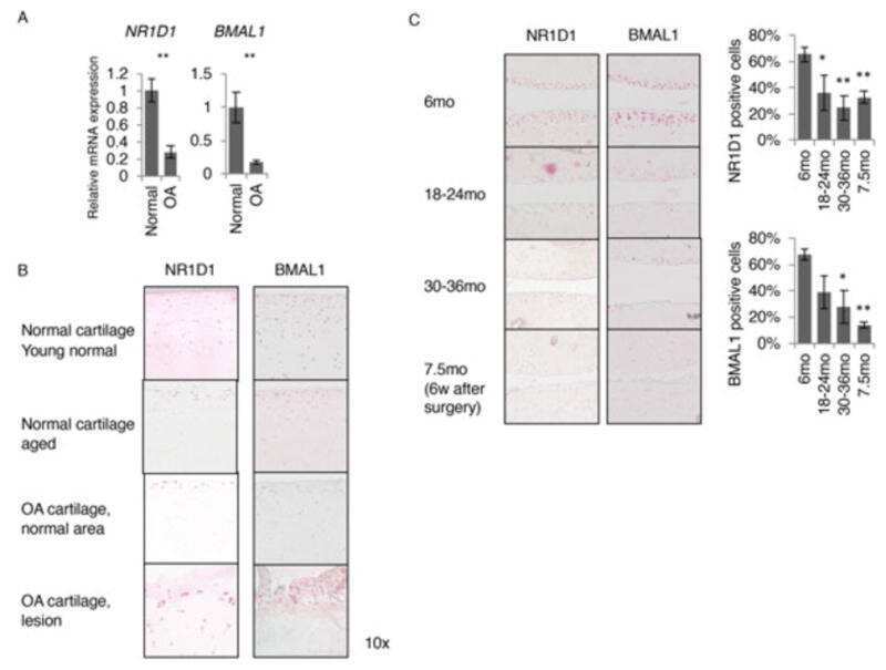
Dysregulated circadian rhythm pathway in human osteoarthritis: NR1D1 and BMAL1 suppression alters TGF-β signaling in chondrocytes.
The transcription factor Runx2 is under circadian control in the suprachiasmatic nucleus and functions in the control of rhythmic behavior.
Expression of circadian rhythm genes in gonadotropin-releasing hormone-secreting GT1-7 neurons.
Circadian Transcription. Thinking outside the E-Box.
Ceci L, Chen L, Baiocchi L, Wu N, Kennedy L, Carpino G, Kyritsi K, Zhou T, Owen T, Kundu D, Sybenga A, Isidan A, Ekser B, Franchitto A, Onori P, Gaudio E, Mancinelli R, Francis H, Alpini G, Glaser S
Cellular and molecular gastroenterology and hepatology 2022;14(4):877-904
Cellular and molecular gastroenterology and hepatology 2022;14(4):877-904
Melatonin modulates daytime-dependent synaptic plasticity and learning efficiency.
Jilg A, Bechstein P, Saade A, Dick M, Li TX, Tosini G, Rami A, Zemmar A, Stehle JH
Journal of pineal research 2019 Apr;66(3):e12553
Journal of pineal research 2019 Apr;66(3):e12553
Dysregulated circadian rhythm pathway in human osteoarthritis: NR1D1 and BMAL1 suppression alters TGF-β signaling in chondrocytes.
Akagi R, Akatsu Y, Fisch KM, Alvarez-Garcia O, Teramura T, Muramatsu Y, Saito M, Sasho T, Su AI, Lotz MK
Osteoarthritis and cartilage 2017 Jun;25(6):943-951
Osteoarthritis and cartilage 2017 Jun;25(6):943-951
The transcription factor Runx2 is under circadian control in the suprachiasmatic nucleus and functions in the control of rhythmic behavior.
Reale ME, Webb IC, Wang X, Baltazar RM, Coolen LM, Lehman MN
PloS one 2013;8(1):e54317
PloS one 2013;8(1):e54317
Expression of circadian rhythm genes in gonadotropin-releasing hormone-secreting GT1-7 neurons.
Gillespie JM, Chan BP, Roy D, Cai F, Belsham DD
Endocrinology 2003 Dec;144(12):5285-92
Endocrinology 2003 Dec;144(12):5285-92
Circadian Transcription. Thinking outside the E-Box.
Muñoz E, Brewer M, Baler R
The Journal of biological chemistry 2002 Sep 27;277(39):36009-17
The Journal of biological chemistry 2002 Sep 27;277(39):36009-17
No comments: Submit comment
Supportive validation
- Submitted by
- Invitrogen Antibodies (provider)
- Main image
- Experimental details
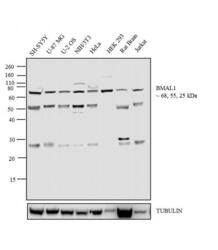
- Western blot analysis was performed on whole cell extracts (30 µg lysate) of SH-SY5Y (Lane 1), U-87 MG (Lane 2), U-2 OS (Lane 3), NIH/3T3 (Lane 4), HeLa (Lane 5), HEK 293 (Lane 6), Rat Brain (tissue extract) (Lane 7) and Jurkat (Lane 8). The blot was probed with Anti-BMAL 1 Rabbit Polyclonal Antibody (Product # PA1-523, 1:100-1:1000 dilution) and detected by chemiluminescence using Goat anti-Rabbit IgG (Heavy Chain) Superclonal™ Secondary Antibody, HRP conjugate (Product # A27036, 0.4 µg/mL, 1:2500 dilution). Three isoforms of 68, 55, 25 kDa corresponding to BMAL 1 were observed across the cell lines and tissue tested. Known quantity of protein samples were electrophoresed using Novex® NuPAGE® 10 % Bis-Tris gel (Product # NP0302BOX), XCell SureLock™ Electrophoresis System (Product # EI0002) and Novex® Sharp Pre-Stained Protein Standard (Product # LC5800). Resolved proteins were then transferred onto a nitrocellulose membrane with iBlot® 2 Dry Blotting System (Product # IB21001). The membrane was probed with the relevant primary and secondary Antibody following blocking with 5 % skimmed milk. Chemiluminescent detection was performed using Pierce™ ECL Western Blotting Substrate (Product # 32106).

- Submitted by
- Invitrogen Antibodies (provider)
- Main image
- Experimental details
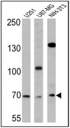
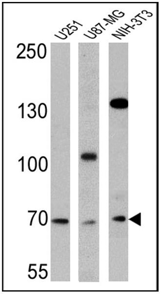
- Western blot analysis of BMAL1 was performed by loading 25 µg of U251 (Lane 1), U87-MG (Lane 2), and NIH-3T3 cell lysates (Lane 3) and a molecular weight protein ladder onto an SDS polyacrylamide gel. Proteins were transferred to a PVDF membrane and blocked with a blocking buffer at 4ºC overnight. The membrane was probed with a BMAL1 polyclonal antibody (Product # PA1-523) at a dilution of 1:500 overnight at 4°C, washed in TBST, and probed with an HRP-conjugated secondary antibody for 1 hr at room temperature in the dark. Chemiluminescent detection was performed using Pierce ECL Plus Western Blotting Substrate (Product # 32132). Results show a band at 69 kDa in all three cell lines.
Supportive validation
- Submitted by
- Invitrogen Antibodies (provider)
- Main image

- Experimental details
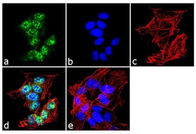
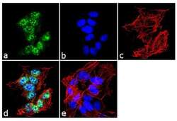
- Immunofluorescent analysis of BMAL1 was performed using 70% confluent log phase SH-SY5Y cells. The cells were fixed with 4% paraformaldehyde for 10 minutes, permeabilized with 0.1% Triton™ X-100 for 10 minutes, and blocked with 1% BSA for 1 hour at room temperature. The cells were labeled with BMAL1 Rabbit Polyclonal Antibody (Product # PA1-523) at 2 µg/mL in 0.1% BSA and incubated for 3 hours at room temperature and then labeled with Goat anti-Rabbit IgG (H+L) Superclonal™ Secondary Antibody, Alexa Fluor® 488 conjugate (Product # A27034) a dilution of 1:2000 for 45 minutes at room temperature (Panel a: green). Nuclei (Panel b: blue) were stained with SlowFade® Gold Antifade Mountant with DAPI (Product # S36938). F-actin (Panel c: red) was stained with Alexa Fluor® 555 Rhodamine Phalloidin (Product # R415, 1:300). Panel d represents the merged image showing nuclear localization. Panel e shows the no primary antibody control. The images were captured at 60X magnification.
- Submitted by
- Invitrogen Antibodies (provider)
- Main image
- Experimental details
- Immunofluorescent analysis of BMAL1 was performed using 70% confluent log phase SH-SY5Y cells. The cells were fixed with 4% paraformaldehyde for 10 minutes, permeabilized with 0.1% Triton™ X-100 for 10 minutes, and blocked with 1% BSA for 1 hour at room temperature. The cells were labeled with BMAL1 Rabbit Polyclonal Antibody (Product # PA1-523) at 2 µg/mL in 0.1% BSA and incubated for 3 hours at room temperature and then labeled with Goat anti-Rabbit IgG (Heavy Chain) Superclonal™ Secondary Antibody, Alexa Fluor® 488 conjugate (Product # A27034) a dilution of 1:2000 for 45 minutes at room temperature (Panel a: green). Nuclei (Panel b: blue) were stained with SlowFade® Gold Antifade Mountant with DAPI (Product # S36938). F-actin (Panel c: red) was stained with Alexa Fluor® 555 Rhodamine Phalloidin (Product # R415, 1:300). Panel d represents the merged image showing nuclear localization. Panel e shows the no primary antibody control. The images were captured at 60X magnification.

Supportive validation
- Submitted by
- Invitrogen Antibodies (provider)
- Main image
- Experimental details
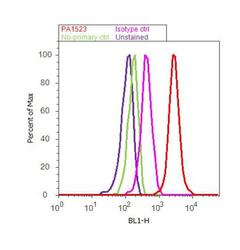
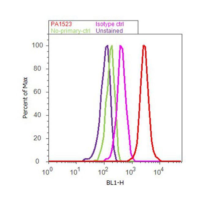
- Flow cytometry analysis of BMAL1 was done on SH-SY5Y cells. Cells were fixed with 70% ethanol for 10 minutes, permeabilized with 0.25% Triton™ X-100 for 20 minutes, and blocked with 5% BSA for 30 minutes at room temperature. Cells were labeled with BMAL1 Rabbit Polyclonal Antibody (PA1-523, red histogram) or with rabbit isotype control (pink histogram) at 3-5 ug/million cells in 2.5% BSA. After incubation at room temperature for 2 hours, the cells were labeled with Alexa Fluor® 488 Goat Anti-Rabbit Secondary Antibody (A11008) at a dilution of 1:400 for 30 minutes at room temperature. The representative 10, 000 cells were acquired and analyzed for each sample using an Attune® Acoustic Focusing Cytometer. The purple histogram represents unstained control cells and the green histogram represents no-primary-antibody control.
Supportive validation
- Submitted by
- Invitrogen Antibodies (provider)
- Main image
- Experimental details
- NULL
- Submitted by
- Invitrogen Antibodies (provider)
- Main image
- Experimental details
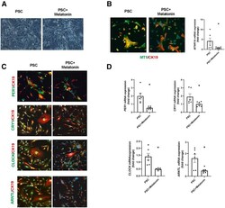
- Melatonin influences the phenotype of isolated P92-PSC. ( A ) Morphologic shapes of human isolated PSC cell line. Scale bars : 20 mum. ( B ) By immunofluorescence and qPCR, the MT1 and ( C and D ) clock genes (PER1, CRY1, CLOCK, and BMAL1) are decreased in isolated PSC cell lines treated with melatonin compared with vehicle-treated PSC cells. Original magnification, 20x; scale bars : 20 mum. Data are means +- SEM of 2 evaluations from 3 cumulative preparations of in vitro experiments. Each dot represents 1 value in data set. * P < .05 vs PSC.
