Explore
 Validate
 Learn
Learn Western blot
Antibody data
- Antibody Data
- Antigen structure
- References [0]
- Comments [0]
- Validations
- Western blot [1]
- Immunocytochemistry [1]
- Immunohistochemistry [2]
- Flow cytometry [1]
Submit
Validation data
Reference
Comment
Report error
- Product number
- AX10006 - Provider product page

- Provider
- Abcepta
- Product name
- ESR2 Antibody (Center)
- Antibody type
- Polyclonal
- Antigen
- Synthetic peptide
- Description
- Peptide Affinity Purified Rabbit Polyclonal Antibody (Pab)
- Reactivity
- Human
- Host
- Rabbit
- Isotype
- IgG
- Vial size
- 400 µl
- Concentration
- 0.5 mg/ml
- Storage
- Maintain refrigerated at 2-8°C for up to 6 months. For long term storage store at -20°C in small aliquots to prevent freeze-thaw cycles.
No comments: Submit comment
Supportive validation
- Submitted by
- Abcepta (provider)
- Main image
- Experimental details
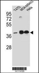
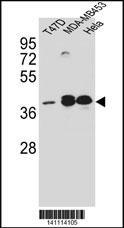
- ESR2 Antibody (Center) (Cat. #AX10006) western blot analysis in T47D ÿMDA-MB453 ÿHela cell line lysates (35ug/lane).This demonstrates the ESR2 antibody detected the ESR2 protein (arrow).
- Primary Ab dilution
- 1:2000
Supportive validation
- Submitted by
- Abcepta (provider)
- Main image
- Experimental details
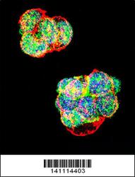
- Confocal immunofluorescent analysis of ESR2 Antibody (Center) with T47D cell followed by Alexa Fluor 488-conjugated goat anti-rabbit lgG (green). Actin filaments have been labeled with Alexa Fluor 555 phalloidin (red). DAPI was used to stain the cell nuclear (blue).
- Primary Ab dilution
- 1:50

Supportive validation
- Submitted by
- Abcepta (provider)
- Main image
- Experimental details
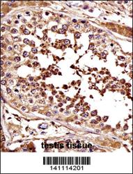
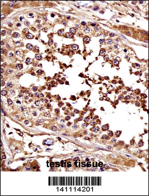
- ESR2 Antibody (Center) immunohistochemistry analysis in formalin fixed and paraffin embedded human testis tissue followed by peroxidase conjugation of the secondary antibody and DAB staining.This data demonstrates the use of ESR2 Antibody (Center) for immunohistochemistry. Clinical relevance has not been evaluated.
- Primary Ab dilution
- 1:50
- Submitted by
- Abcepta (provider)
- Main image
- Experimental details
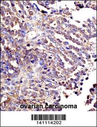
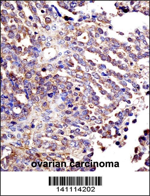
- ESR2 Antibody (Center) immunohistochemistry analysis in formalin fixed and paraffin embedded human ovarian carcinoma followed by peroxidase conjugation of the secondary antibody and DAB staining.This data demonstrates the use of ESR2 Antibody (Center) for immunohistochemistry. Clinical relevance has not been evaluated.
- Primary Ab dilution
- 1:50
Supportive validation
- Submitted by
- Abcepta (provider)
- Main image
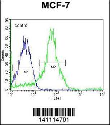
- Experimental details
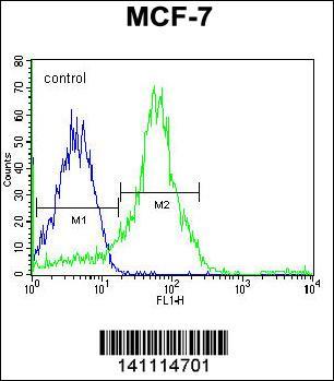
- ESR2 Antibody (Center) flow cytometric analysis of MCF-7 cells (right histogram) compared to a negative control cell (left histogram).Alexa Fluor 488-conjugated donkey anti-rabbit lgG secondary antibodies were used for the analysis.
- Primary Ab dilution
- 1:50