Explore
 Validate
 Learn
Learn Western blot
 Immunohistochemistry
Antibody data
- Antibody Data
- Antigen structure
- References [0]
- Comments [0]
- Validations
- Immunohistochemistry [4]
- Flow cytometry [4]
Submit
Validation data
Reference
Comment
Report error
- Product number
- NBP2-44366-0.1mg - Provider product page

- Provider
- Novus Biologicals
- Product name
- Mouse Monoclonal ER beta/NR3A2 Antibody
- Antibody type
- Monoclonal
- Description
- Protein A or G purified. Estrogen receptors (ER) are members of the steroid/thyroid hormone receptor superfamily of ligand-activated transcription factors. Estrogen receptors, including ER-alpha and ER-beta, contain DNA binding and ligand binding domains and are critically involved in regulating the normal function of reproductive tissues. They are located in the nucleus, though some estrogen receptors associate with the cell surface membrane and can be rapidly activated by exposure of cells to estrogen. ER-alpha and ER-beta are differentially activated by various ligands. Receptor-ligand interactions trigger a cascade of events, including dissociation from heat shock proteins, receptor dimerization, phosphorylation and the association of the hormone activated receptor with specific regulatory elements in target genes. Evidence suggests that ER-alpha and ER-beta may be regulated by distinct mechanisms even though they share many functional characteristics.
- Reactivity
- Human
- Host
- Mouse
- Isotype
- IgG
- Vial size
- 0.1 mg
- Concentration
- 0.2 mg/ml
- Storage
- Store at 4C.
No comments: Submit comment
Supportive validation
- Submitted by
- Novus Biologicals (provider)
- Main image

- Experimental details
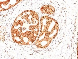
- Immunohistochemistry-Paraffin: ER beta/NR3A2 Antibody (ESR2/686) [NBP2-44366] - Human Bladder Carcinoma stained with ER-beta1 Monoclonal Antibody (ESR2/686).

- Submitted by
- Novus Biologicals (provider)
- Main image
- Experimental details
- Immunohistochemistry-Paraffin: ER beta/NR3A2 Antibody (ESR2/686) [NBP2-44366] - Human Gastric Carcinoma stained with ER-beta1 Monoclonal Antibody (ESR2/686).

- Submitted by
- Novus Biologicals (provider)
- Main image
- Experimental details
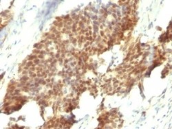
- Immunohistochemistry-Paraffin: ER beta/NR3A2 Antibody (ESR2/686) [NBP2-44366] - Human Breast Carcinoma stained with ER-beta1 Monoclonal Antibody (ESR2/686).

- Submitted by
- Novus Biologicals (provider)
- Main image

- Experimental details
- Immunohistochemistry-Paraffin: ER beta/NR3A2 Antibody (ESR2/686) [NBP2-44366] - Human Ovarian Carcinoma stained with ER-beta1 Monoclonal Antibody (ESR2/686).

Supportive validation
- Submitted by
- Novus Biologicals (provider)
- Main image

- Experimental details
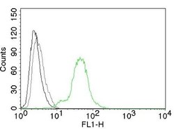
- Flow Cytometry: ER beta/NR3A2 Antibody (ESR2/686) [NBP2-44366] - Human ER beta on BT474 Cells. Black: Cells alone; Grey: Isotype Control; Green: AF488-labeled ER beta1 Monoclonal Antibody (ESR2/686).

- Submitted by
- Novus Biologicals (provider)
- Main image

- Experimental details
- Flow Cytometry: ER beta/NR3A2 Antibody (ESR2/686) [NBP2-44366] - Flow Cytometry: ER beta/NR3A2 Antibody (ESR2/686) [Phycoerythrin] [NBP2-47797PE] - An intracellular stain was performed on MCF-7 cells with ER Beta/NR3A2 (ESR2/686) NBP2-47797PE (blue) and a matched isotype control (orange). Cells were fixed with 4% PFA and then permeablized with 0.1% saponin. Cells were incubated in an antibody dilution of 1 ug/mL for 30 minutes at room temperature. Both antibodies were conjugated to phycoerythrin. Image using the PE form of this antibody.

- Submitted by
- Novus Biologicals (provider)
- Main image
- Experimental details
- Flow Cytometry: ER beta/NR3A2 Antibody (ESR2/686) [NBP2-44366] - Flow Cytometry of human ER beta on BT474 cells. Black: cells alone; Grey: Isotype Control; Green: AF488-labeled ER beta/NR3A2 Antibody (ESR2/686).

- Submitted by
- Novus Biologicals (provider)
- Main image
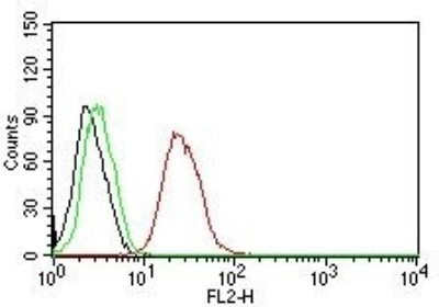
- Experimental details
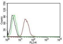
- Flow Cytometry: ER beta/NR3A2 Antibody (ESR2/686) [NBP2-44366] - Flow Cytometry for human ER-beta on MCF-7 cells. Black: cells alone; Green: Isotype Control; Red: PE-labeled ER beta/NR3A2 Antibody (ESR2/686).