Explore
 Validate
 Learn
Learn Western blot
 Immunocytochemistry
Antibody data
- Antibody Data
- Antigen structure
- References [1]
- Comments [0]
- Validations
- Immunocytochemistry [2]
- Immunohistochemistry [2]
- Other assay [2]
Submit
Validation data
Reference
Comment
Report error
- Product number
- PA5-104479 - Provider product page

- Provider
- Invitrogen Antibodies
- Product name
- LIGHT Polyclonal Antibody
- Antibody type
- Polyclonal
- Antigen
- Synthetic peptide
- Description
- Antibody detects endogenous levels of total TNFSF14.
- Reactivity
- Human, Mouse
- Host
- Rabbit
- Isotype
- IgG
- Vial size
- 100 μL
- Concentration
- 1 mg/mL
- Storage
- -20°C
Submitted references HES5-mediated repression of LIGHT transcription may contribute to apoptosis in hepatocytes.
Miao X, Guo Y, Zeng S, Liu X, Teng X, Li L, Hong W
Cell death discovery 2021 Oct 23;7(1):308
Cell death discovery 2021 Oct 23;7(1):308
No comments: Submit comment
Supportive validation
- Submitted by
- Invitrogen Antibodies (provider)
- Main image
- Experimental details
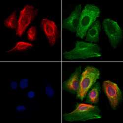
- Immunofluorescent analysis of LIGHT in HeLa cells. Samples were fixed with paraformaldehyde, permeabilized with 0.1% Triton X-100, blocked with 10% serum (45 min at 25°C), incubated with mouse anti-beta tubulin and LIGHT polyclonal antibody (Product # PA5-104479) using a dilution of 1:200 (1 hr, 37°C), and followed by goat anti-rabbit IgG Alexa Fluor 594 (red) and goat anti-mouse IgG Alexa Fluor 488 (green).
- Submitted by
- Invitrogen Antibodies (provider)
- Main image

- Experimental details
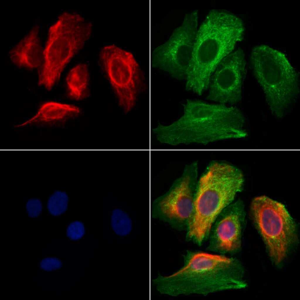
- Immunofluorescent analysis of LIGHT in HeLa cells. Samples were fixed with paraformaldehyde, permeabilized with 0.1% Triton X-100, blocked with 10% serum (45 min at 25°C), incubated with mouse anti-beta tubulin and LIGHT polyclonal antibody (Product # PA5-104479) using a dilution of 1:200 (1 hr, 37°C), and followed by goat anti-rabbit IgG Alexa Fluor 594 (red) and goat anti-mouse IgG Alexa Fluor 488 (green).
Supportive validation
- Submitted by
- Invitrogen Antibodies (provider)
- Main image

- Experimental details
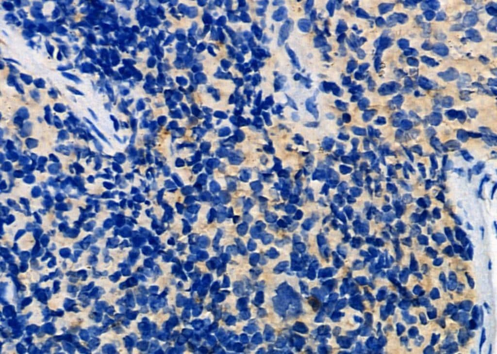
- Immunohistochemistry analysis of LIGHT in mouse spleen tissue. The sample was formaldehyde fixed and a heat mediated antigen retrieval step in citrate buffer was performed. Samples were incubated with LIGHT polyclonal antibody (Product # PA5-104479) using a dilution of 1:100 (4°C overnight) followed by HRP conjugated anti-Rabbit secondary antibody.
- Submitted by
- Invitrogen Antibodies (provider)
- Main image
- Experimental details
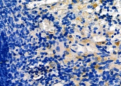
- Immunohistochemistry analysis of LIGHT in rat spleen tissue. The sample was formaldehyde fixed and a heat mediated antigen retrieval step in citrate buffer was performed. Samples were incubated with LIGHT polyclonal antibody (Product # PA5-104479) using a dilution of 1:100 (4°C overnight) followed by HRP conjugated anti-Rabbit secondary antibody.

Supportive validation
- Submitted by
- Invitrogen Antibodies (provider)
- Main image
- Experimental details
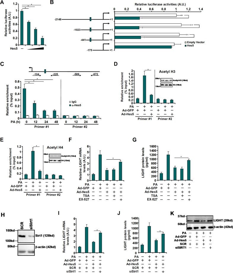
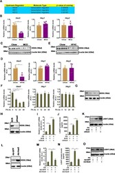
- Fig. 3 HES5 downregulation parallels LIGHT upregulation in the pathogenesis of non-alcoholic fatty liver disease. A IPA analysis of potential LIGHT upstream regulators. B , C C57B6/L mice were fed an MCD diet for 4 weeks. Hepatic gene expression was examined by qPCR and western. D , E C57B6/L mice were fed an HFHC diet for 12 weeks. Hepatic gene expression was examined by qPCR and western. F , G Primary murine hepatocytes were treated with palmitate (0.3 mM) and harvested at indicated time points. Gene expression was examined by qPCR and ELISA. H - K Primary murine hepatocytes were transfected with siRNA-targeting LIGHT or scrambled siRNA (SCR) followed by treatment with PA (0.3 mM). LIGHT expression was examined by qPCR, ELISA, and western blotting. L - O Primary murine hepatocytes were transduced with Ad-HES5 or Ad-GFP followed by treatment with PA (0.3 mM). LIGHT expression was examined by qPCR, ELISA, and western blotting.
- Submitted by
- Invitrogen Antibodies (provider)
- Main image
- Experimental details
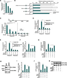
- Fig. 4 HES5 directly binds to the LIGHT promoter and represses LIGHT transcription. A A LIGHT promoter-luciferase construct (-2186/+1) were transfected into HepG2 cells with or without HEY5. Luciferase activities were normalized by protein concentration and GFP fluorescence. B Different LIGHT promoter-luciferase constructs were transfected into HepG2 cells with or without HES5. Luciferase activities were normalized by protein concentration and GFP fluorescence. C Primary murine hepatocytes were treated with palmitate (0.3 mM) and harvested at indicated time points. ChIP assay was performed with anti-HES5 or IgG. D , E Primary murine hepatocytes were transduced with Ad-HES5 or Ad-GFP followed by treatment with PA (0.3 mM). ChIP assay was performed with anti-acetyl H3 and anti-acetyl H4. Inset, global histone H3/H4 and acetyl H3/H4 levels were examined by western blotting. F , G Primary murine hepatocytes were transduced with Ad-HES5 or Ad-GFP followed by treatment with PA (0.3 mM) in the presence of absence of TSA (100 nM) or EX-527 (1 muM). LIGHT expression was examined by qPCR and ELISA. H-K Primary murine hepatocytes were transduced with Ad-HES5 or Ad-GFP, transfected with siRNA-targeting SIRT1 or scrambled siRNA (SCR) and treated with PA (0.3 mM). SIRT1 knockdown efficiency was examined by western. LIGHT expression was examined by qPCR, ELISA, and western blotting.