Explore
 Validate
 Learn
Learn Western blot
 Immunocytochemistry
Antibody data
- Antibody Data
- Antigen structure
- References [1]
- Comments [0]
- Validations
- Immunocytochemistry [6]
- Other assay [1]
Submit
Validation data
Reference
Comment
Report error
- Product number
- MA5-32406 - Provider product page

- Provider
- Invitrogen Antibodies
- Product name
- APC6 Recombinant Rabbit Monoclonal Antibody (SD085-8)
- Antibody type
- Monoclonal
- Antigen
- Recombinant full-length protein
- Description
- Recombinant rabbit monoclonal antibodies are produced using in vitro expression systems. The expression systems are developed by cloning in the specific antibody DNA sequences from immunoreactive rabbits. Then, individual clones are screened to select the best candidates for production. The advantages of using recombinant rabbit monoclonal antibodies include: better specificity and sensitivity, lot-to-lot consistency, animal origin-free formulations, and broader immunoreactivity to diverse targets due to larger rabbit immune repertoire.
- Reactivity
- Human, Mouse, Rat
- Host
- Rabbit
- Isotype
- IgG
- Antibody clone number
- SD085-8
- Vial size
- 100 μL
- Concentration
- 1 mg/mL
- Storage
- Store at 4°C short term. For long term storage, store at -20°C, avoiding freeze/thaw cycles.
Submitted references Downregulated genes by silencing MYC pathway identified with RNA-SEQ analysis as potential prognostic biomarkers in gastric adenocarcinoma.
Heitor da Silva Maués J, Ferreira Ribeiro H, de Maria Maués Sacramento R, Maia de Sousa R, Pereira de Tommaso R, Dourado Kovacs Machado Costa B, Cardoso Soares P, Pimentel Assumpção P, de Fátima Aquino Moreira-Nunes C, Mário Rodriguez Burbano R
Aging 2020 Dec 22;12(24):24651-24670
Aging 2020 Dec 22;12(24):24651-24670
No comments: Submit comment
Supportive validation
- Submitted by
- Invitrogen Antibodies (provider)
- Main image

- Experimental details
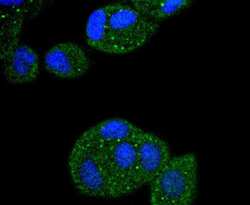
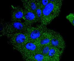
- Immunocytochemical analysis of APC6 in HepG2 cells using a APC6 Monoclonal antibody (Product # MA5-32406) as seen in green. The nuclear counter stain is DAPI (blue). Cells were fixed in paraformaldehyde, permeabilised with 0.25% Triton X100/PBS.

- Submitted by
- Invitrogen Antibodies (provider)
- Main image
- Experimental details
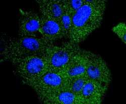
- Immunocytochemical analysis of APC6 in MCF-7 cells using a APC6 Monoclonal antibody (Product # MA5-32406) as seen in green. The nuclear counter stain is DAPI (blue). Cells were fixed in paraformaldehyde, permeabilised with 0.25% Triton X100/PBS.

- Submitted by
- Invitrogen Antibodies (provider)
- Main image
- Experimental details
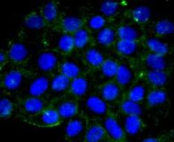
- Immunocytochemical analysis of APC6 in RH-35 cells using a APC6 Monoclonal antibody (Product # MA5-32406) as seen in green. The nuclear counter stain is DAPI (blue). Cells were fixed in paraformaldehyde, permeabilised with 0.25% Triton X100/PBS.

- Submitted by
- Invitrogen Antibodies (provider)
- Main image
- Experimental details
- Immunocytochemical analysis of APC6 in HepG2 cells using a APC6 Monoclonal antibody (Product # MA5-32406) as seen in green. The nuclear counter stain is DAPI (blue). Cells were fixed in paraformaldehyde, permeabilised with 0.25% Triton X100/PBS.

- Submitted by
- Invitrogen Antibodies (provider)
- Main image

- Experimental details
- Immunocytochemical analysis of APC6 in MCF-7 cells using a APC6 Monoclonal antibody (Product # MA5-32406) as seen in green. The nuclear counter stain is DAPI (blue). Cells were fixed in paraformaldehyde, permeabilised with 0.25% Triton X100/PBS.

- Submitted by
- Invitrogen Antibodies (provider)
- Main image
- Experimental details
- Immunocytochemical analysis of APC6 in RH-35 cells using a APC6 Monoclonal antibody (Product # MA5-32406) as seen in green. The nuclear counter stain is DAPI (blue). Cells were fixed in paraformaldehyde, permeabilised with 0.25% Triton X100/PBS.

Supportive validation
- Submitted by
- Invitrogen Antibodies (provider)
- Main image
- Experimental details
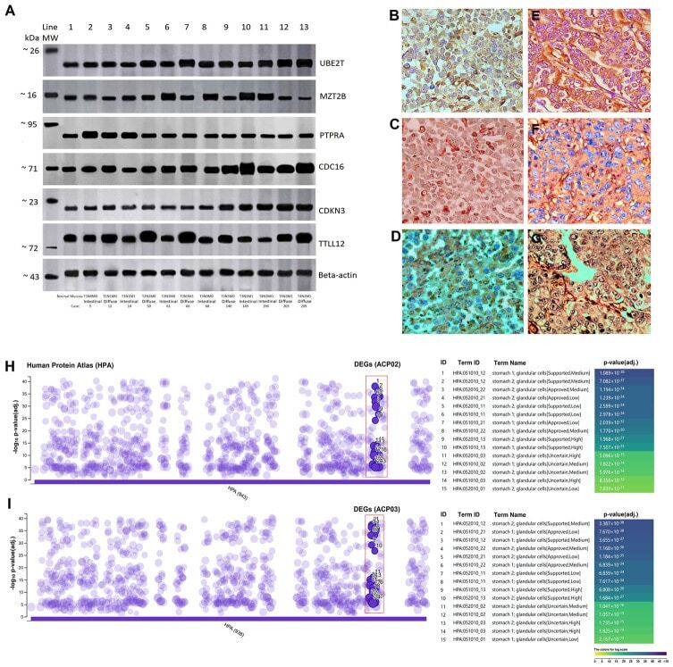
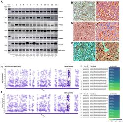
- Figure 3 Analysis of protein expression and immunohistochemistry in gastric cancer subtypes. ( A ) Representative image of Western-blot. Line 1 represents normal gastric tissue and the intensity of the genes are similar to early TNM stages and without metastasis, but much lower than advanced TNM stages (T3/T4). Initial stages with metastasis also have an intensity much higher than that of normal gastric tissue. ( B ) Positive TTLL12 cytoplasmatic immunostaining in diffuse-type gastric cancer (case 66 T2N3M1); ( C ) Positive CDC16 cytoplasmic and nuclear immunostaining in diffuse-type gastric cancer (case 140 T3N3M0); ( D ) Positive CDKN3 cytoplasmatic immunostaining in diffuse-type gastric cancer (case 203 T4N2M1); ( E ) Positive PTPRA cytoplasmatic immunostaining in intestinal-type gastric cancer (case 5 T1N0M0); ( F ) Positive MZT2B cytoplasmatic immunostaining in intestinal-type gastric cancer (case 61 T2N3M0); ( G ) Positive UBE2T cytoplasmatic and nuclear immunostaining in intestinal-type gastric cancer (case 149 T3N3M1) (magnification x40). The differences in band intensity and intensity of immunoreactivity are due to the different stages of TNM in tumor samples of diffuse and intestinal histological types that represent figures ( 3A , 3B - 3G ). ( H and I ) The function of the DEGs in the gastric lines showed a strong correlation between the increased level of protein expression with the human stomach cells in the data from The Human Protein Atlas (HPA) that were acces