 Explore
 Validate
 Learn
Learn Western blot
 Immunohistochemistry
Antibody data
- Antibody Data
- Antigen structure
- References [1]
- Comments [0]
- Validations
- Immunohistochemistry [1]
Submit
Validation data
Reference
Comment
Report error
- Product number
- MA3-814 - Provider product page

- Provider
- Invitrogen Antibodies
- Product name
- S-arrestin Monoclonal Antibody (PDS1)
- Antibody type
- Monoclonal
- Antigen
- Other
- Description
- MA3-814 detects Visual Arrestin from human, rat, porcine, bovine, ovine, and goat tissues. MA3-814 has been successfully used in Immunohistochemistry, ELISA, and Western blotting procedures. By Western blot it detects a 37-kDa band representing Visual arrestin. The MA3-814 antigen is Visual arrestin isolated from Porcine cells.
- Reactivity
- Human, Rat, Bovine, Goat, Porcine
- Host
- Mouse
- Isotype
- IgG
- Antibody clone number
- PDS1
- Vial size
- 100 μL
- Concentration
- Conc. Not Determined
- Storage
- -20°C, Avoid Freeze/Thaw Cycles
Submitted references Analysis of antigenic determinants of retinal S-antigen with monoclonal antibodies.
Banga JP, LeRoy F, Suleyman S, Kasp E, Brown E, Dumonde D
Investigative ophthalmology & visual science 1988 Jan;29(1):12-21
Investigative ophthalmology & visual science 1988 Jan;29(1):12-21
No comments: Submit comment
Supportive validation
- Submitted by
- Invitrogen Antibodies (provider)
- Main image
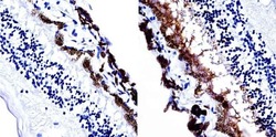
- Experimental details
- Immunohistochemistry was performed on human retina tissue. To expose target protein, antigen was retreived using 10mM sodium citrate followed by microwave treatment for 8-15 minutes. Endogenous peroxidases were blocked in 3% H202-methanol for 15 minutes and tissues were blocked in 3% BSA-PBS for 30 minutes at room temperature. Cells were probed with a Visual Arrestin mouse monoclonal antibody (Product # MA3-814) at a dilution of 1:20 overnight in a humidified chamber. Tissues were washed in PBST and detection was performed using a secondary antibody conjugated to HRP. DAB staining buffer was applied and tissues were counterstained with hematoxylin and prepped for mounting. Images were taken at 40X magnification.
