Explore
 Validate
 Learn
Learn Western blot
Antibody data
- Antibody Data
- Antigen structure
- References [1]
- Comments [0]
- Validations
- Western blot [2]
- Immunohistochemistry [1]
Submit
Validation data
Reference
Comment
Report error
- Product number
- 14-9961-82 - Provider product page

- Provider
- Invitrogen Antibodies
- Product name
- MALT1 Monoclonal Antibody (50), eBioscience™
- Antibody type
- Monoclonal
- Antigen
- Other
- Description
- Description: The monoclonal antibody 50 recognizes human MALT1 (Mucosal Associated Lymphoid Tissue lymphoma translocation gene 1). MALT1 is a 91 kDa paracaspase that contains a death domain, two Ig-like domains and a C terminal caspase-like domain. Like other caspases, it cleaves substrates after an arginine residue. Expression is found in the cytoplasm of B and T cells. Within B cell follicles, MALT1 expression is highly expressed in centroblasts, followed by centrocytes and weakly expressed in the mantle zone. The interaction of MALT1 and Bcl10 occurs through Ig-like domains resulting in oligomerization and activation of the caspase-like domain. This also directly results in NFkappa B activation thereby playing a role in B cell maturation and activation.
- Antibody clone number
- 50
- Concentration
- 0.5 mg/mL
Submitted references A novel fusion of the MALT1 gene and the microtubule-associated protein 4 (MAP4) gene occurs in diffuse large B-cell lymphoma.
Murga Penas EM, Kawadler H, Siebert R, Frank M, Ye H, Hinz K, Becher C, Hummel M, Barth TF, Bokemeyer C, Stein H, Trümper L, Möller P, Marynen P, Du MQ, Yang X, Hansmann ML, Dierlamm J
Genes, chromosomes & cancer 2006 Sep;45(9):863-73
Genes, chromosomes & cancer 2006 Sep;45(9):863-73
No comments: Submit comment
Supportive validation
- Submitted by
- Invitrogen Antibodies (provider)
- Main image
- Experimental details
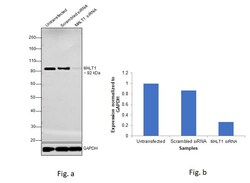
- Knockdown of MALT1 was achieved by transfecting HeLa with MALT1 specific siRNAs (Silencer® select Product # s21398). Western blot analysis (Fig. a) was performed using whole cell extracts from the MALT1 knockdown cells (lane 3), non-specific scrambled siRNA transfected cells (lane 2) and untransfected cells (lane 1). The blot was probed with MALT1 Monoclonal Antibody (Product # 14-9961-82, 5µg/ml) and Goat anti-Mouse IgG (H+L), Superclonal™ Recombinant Secondary Antibody, HRP (Product # A28177, 1:4000 dilution). Densitometric analysis of this western blot is shown in histogram (Fig. b). Decrease in signal upon siRNA mediated knock down confirms that antibody is specific to MALT1.

- Submitted by
- Invitrogen Antibodies (provider)
- Main image
- Experimental details
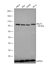
- Western blot was performed using Anti-MALT1 Monoclonal Antibody (Product # 14-9961-82) and a 92 kDa band corresponding to MALT1 was observed in Hela, Raji, Jurkat and PC-3. Whole cell extracts (30 µg lysate) of HeLa (Lane 1), Raji (Lane 2), Jurkat (Lane 3) and PC-3 (Lane 4) were electrophoresed using Novex® NuPAGE® 4-12 % Bis-Tris gel (Product # NP0322BOX). Resolved proteins were then transferred onto a nitrocellulose membrane (Product # IB23001) by iBlot® 2 Dry Blotting System (Product # IB21001). The blot was probed with the primary antibody (5µg/ml) and detected by chemiluminescence with Goat anti-Mouse IgG (H+L), Superclonal™ Recombinant Secondary Antibody, HRP (Product # A28177, 1:4000 dilution) using the iBright FL 1000 (Product # A32752). Chemiluminescent detection was performed using Novex® ECL Chemiluminescent Substrate Reagent Kit (Product # WP20005).
Supportive validation
- Submitted by
- Invitrogen Antibodies (provider)
- Main image
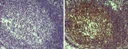
- Experimental details
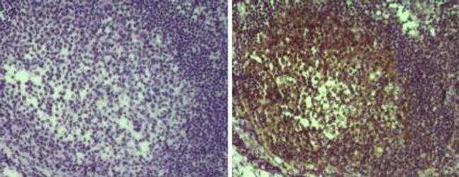
- Immunohistochemistry on formalin-fixed paraffin embedded human tonsil tissue with citrate buffer antigen retrieval, using 10 µg/mL of Anti-Human MALT1 Purified (right) or Mouse IgG1 kappa Isotype Control Purified (Product # 14-4714-82) antibody (left) followed by Anti-Mouse Ig Biotin and DAB visualization. Nuclei are counterstained with hematoxylin.