 Explore
 Validate
 Learn
Learn Western blot
 ELISA
ELISAAntibody data
- Antibody Data
- Antigen structure
- References [0]
- Comments [0]
- Validations
- Western blot [1]
- Immunohistochemistry [2]
Submit
Validation data
Reference
Comment
Report error
- Product number
- LS-C154236 - Provider product page

- Provider
- LSBio
- Product name
- ABCB5 Antibody (Internal) LS-C154236
- Antibody type
- Polyclonal
- Description
- Affinity purified
- Reactivity
- Human
- Host
- Rabbit
- Isotype
- IgG
- Storage
- Short term: store at 4°C. Long term: aliquot and store at -20°C. Avoid freeze-thaw cycles.
No comments: Submit comment
Enhanced validation
- Submitted by
- LSBio (provider)
- Enhanced method
- Genetic validation
- Main image
- Experimental details
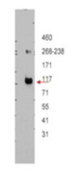
- Western Blot - ABCB5 Antibody. Western blot of affinity purified anti-ABCB5 antibody shows detection of ABCB5 beta in ~12.5 ug of transfected-Hi5 whole cell lysate. No reaction was seen when antibody was pre-incubated with the immunizing peptide (data not shown). A 3-8% Tris-acetate gel was used for separation. The arrowhead corresponds to 117 kD ABCB5. The membrane was probed with the primary antibody at a 1:10000 dilution in 5% milk in TBST at 4C, overnight. Personal Communication, JP Gillet, CCR-NCI, Bethesda, MD.

Enhanced validation
- Submitted by
- LSBio (provider)
- Enhanced method
- Genetic validation
- Main image

- Experimental details
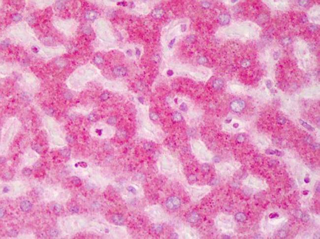
- IHC - ABCB5 Antibody. Immunohistochemistry of ABCB5 antibody tissue: human liver Fixation: formalin fixed paraffin embedded Antigen retrieval: user optimized Primary antibody: ABCB5 1:200 Secondary antibody: Peroxidase goat anti-rabbit at 1:10000 for 45 min at RT Localization: Moderate to strong cytoplasmic and membranous staining was observed in hepatocytes. Occasional nuclear staining was observed in hepatocytes and sinusoidal cells. Staining: antibody as precipitated red signal with a hematoxylin purple nuclear counterstain.
- Submitted by
- LSBio (provider)
- Main image
- Experimental details
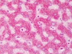
- IHC - ABCB5 Antibody. Immunohistochemistry of ABCB5 antibody tissue: human liver Fixation: formalin fixed paraffin embedded Antigen retrieval: user optimized Primary antibody: ABCB5 1:200 Secondary antibody: Peroxidase goat anti-rabbit at 1:10000 for 45 min at RT Localization: Moderate to strong cytoplasmic and membranous staining was observed in hepatocytes. Occasional nuclear staining was observed in hepatocytes and sinusoidal cells. Staining: antibody as precipitated red signal with a hematoxylin purple nuclear counterstain.
