 Explore
 Validate
 Learn
Learn Western blot
 ELISA
ELISA Immunohistochemistry
Antibody data
- Antibody Data
- Antigen structure
- References [0]
- Comments [0]
- Validations
- Immunohistochemistry [6]
Submit
Validation data
Reference
Comment
Report error
- Product number
- LS-C745315 - Provider product page

- Provider
- LSBio
- Product name
- ABCB5 Antibody (Internal) LS-C745315
- Antibody type
- Polyclonal
- Description
- Affinity purified
- Reactivity
- Human
- Host
- Rabbit
- Isotype
- IgG
- Storage
- Store vial at -20°C or below prior to opening. Dilute 1:10 to minimize loss. Store the vial at -20°C or below after dilution. Avoid freeze-thaw cycles.
No comments: Submit comment
Supportive validation
- Submitted by
- LSBio (provider)
- Enhanced method
- Genetic validation
- Main image
- Experimental details
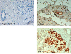
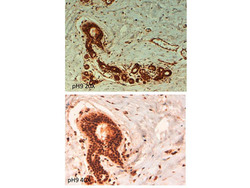
- Immunohistochemistry of rabbit anti ABCB5 antibody. Tissue: human breast carcinoma at pH6. Fixation: formalin fixed paraffin embedded. Primary antibody: ABCB5 antibody at 10 µg/mL for 1 h at RT. Secondary antibody: Peroxidase rabbit secondary antibody at 1:10,000 for 45 min at RT. Localization: ABCB5 is cytoplasmic. Staining: ABCB5 as precipitated brown signal with hematoxylin purple nuclear counterstain.

- Submitted by
- LSBio (provider)
- Enhanced method
- Genetic validation
- Main image
- Experimental details
- Immunohistochemistry of rabbit anti ABCB5 antibody. Tissue: human breast carcinoma at pH9. Fixation: formalin fixed paraffin embedded. Primary antibody: ABCB5 antibody at 10 µg/mL for 1 h at RT. Secondary antibody: Peroxidase rabbit secondary antibody at 1:10,000 for 45 min at RT. Localization: ABCB5 is cytoplasmic. Staining: ABCB5 as precipitated brown signal.

- Submitted by
- LSBio (provider)
- Enhanced method
- Genetic validation
- Main image
- Experimental details
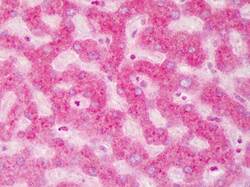
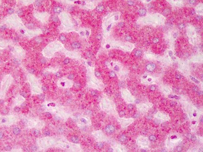
- Immunohistochemistry of ABCB5 antibody Tissue: human liver Fixation: formalin fixed paraffin embedded Antigen retrieval: user optimized Primary antibody: ABCB5 1:200 Secondary antibody: Peroxidase goat anti-rabbit at 1:10,000 for 45 min at RT Localization: Moderate to strong cytoplasmic and membranous staining was observed in hepatocytes. Occasional nuclear staining was observed in hepatocytes and sinusoidal cells. Staining: antibody as precipitated red signal with a hematoxylin purple nuclear counterstain
- Submitted by
- LSBio (provider)
- Main image

- Experimental details
- Immunohistochemistry of rabbit anti ABCB5 antibody. Tissue: human breast carcinoma at pH6. Fixation: formalin fixed paraffin embedded. Primary antibody: ABCB5 antibody at 10 µg/mL for 1 h at RT. Secondary antibody: Peroxidase rabbit secondary antibody at 1:10,000 for 45 min at RT. Localization: ABCB5 is cytoplasmic. Staining: ABCB5 as precipitated brown signal with hematoxylin purple nuclear counterstain.

- Submitted by
- LSBio (provider)
- Main image

- Experimental details
- Immunohistochemistry of rabbit anti ABCB5 antibody. Tissue: human breast carcinoma at pH9. Fixation: formalin fixed paraffin embedded. Primary antibody: ABCB5 antibody at 10 µg/mL for 1 h at RT. Secondary antibody: Peroxidase rabbit secondary antibody at 1:10,000 for 45 min at RT. Localization: ABCB5 is cytoplasmic. Staining: ABCB5 as precipitated brown signal.

- Submitted by
- LSBio (provider)
- Main image

- Experimental details
- Immunohistochemistry of ABCB5 antibody Tissue: human liver Fixation: formalin fixed paraffin embedded Antigen retrieval: user optimized Primary antibody: ABCB5 1:200 Secondary antibody: Peroxidase goat anti-rabbit at 1:10,000 for 45 min at RT Localization: Moderate to strong cytoplasmic and membranous staining was observed in hepatocytes. Occasional nuclear staining was observed in hepatocytes and sinusoidal cells. Staining: antibody as precipitated red signal with a hematoxylin purple nuclear counterstain
