Explore
 Validate
 Learn
Learn Western blot
 Immunohistochemistry
Antibody data
- Antibody Data
- Antigen structure
- References [3]
- Comments [0]
- Validations
- Immunohistochemistry [1]
- Other assay [5]
Submit
Validation data
Reference
Comment
Report error
- Product number
- 14-9758-82 - Provider product page

- Provider
- Invitrogen Antibodies
- Product name
- HGAL Monoclonal Antibody (1H1-A7), eBioscience™
- Antibody type
- Monoclonal
- Antigen
- Other
- Description
- Description: The monoclonal antibody 1H1-A7 recognizes human germinal center-associated lymphoma (HGAL) protein. HGAL is expressed in germinal centers, spleen, thymus, and germinal center-derived lymphomas. HGAL is localized to the cytoplasm of cells within the dark zone of the germinal center and functions to enhance the activation of RhoA leading to negative regulation of lymphocyte motility. HGAL expression has been used to help elucidate nodal marginal zone lymphoma (NMZL) from cases of diffuse follicle center lymphoma. Additionally, HGAL expression was shown to correlate with survival in patients with diffuse large B-cell lymphoma (DLBCL). The 1H1-A7 antibody recognizes the protein product of the M17 gene, the mouse homolog of the HGAL protein. Applications Reported: This 1H1-A7 antibody has been reported for use in immunoprecipitation, western blotting, immunohistochemical staining of formalin-fixed paraffin embedded tissue sections, microscopy, and ELISA. Applications Tested: This 1H1-A7 antibody has been tested by immunohistochemistry of formalin-fixed paraffin embedded human tissue using low pH antigen retrieval and can be used at less than or equal to 5 µg/mL. This 1H1-A7 antibody has been tested by western blot of reduced human and mouse lysate and can be used at less than or equal to 5 µg/mL. Iti s recommended that the antibody be carefully titrated for optimal performance in the assay of interest. Purity: Greater than 90%, as determined by SDS-PAGE. Aggregation: Less than 10%, as determined by HPLC. Filtration: 0.2 µm post-manufacturing filtered.
- Reactivity
- Human, Mouse
- Host
- Mouse
- Isotype
- IgG
- Antibody clone number
- 1H1-A7
- Vial size
- 100 μg
- Concentration
- 0.5 mg/mL
- Storage
- 4°C
Submitted references Targeting N-myristoylation for therapy of B-cell lymphomas.
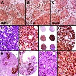
Immunoarchitectural patterns in nodal marginal zone B-cell lymphoma: a study of 51 cases.
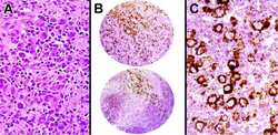
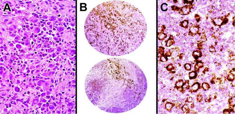
Expression of the human germinal center-associated lymphoma (HGAL) protein, a new marker of germinal center B-cell derivation.
Beauchamp E, Yap MC, Iyer A, Perinpanayagam MA, Gamma JM, Vincent KM, Lakshmanan M, Raju A, Tergaonkar V, Tan SY, Lim ST, Dong WF, Postovit LM, Read KD, Gray DW, Wyatt PG, Mackey JR, Berthiaume LG
Nature communications 2020 Oct 22;11(1):5348
Nature communications 2020 Oct 22;11(1):5348
Immunoarchitectural patterns in nodal marginal zone B-cell lymphoma: a study of 51 cases.
Salama ME, Lossos IS, Warnke RA, Natkunam Y
American journal of clinical pathology 2009 Jul;132(1):39-49
American journal of clinical pathology 2009 Jul;132(1):39-49
Expression of the human germinal center-associated lymphoma (HGAL) protein, a new marker of germinal center B-cell derivation.
Natkunam Y, Lossos IS, Taidi B, Zhao S, Lu X, Ding F, Hammer AS, Marafioti T, Byrne GE Jr, Levy S, Warnke RA, Levy R
Blood 2005 May 15;105(10):3979-86
Blood 2005 May 15;105(10):3979-86
No comments: Submit comment
Supportive validation
- Submitted by
- Invitrogen Antibodies (provider)
- Main image
- Experimental details
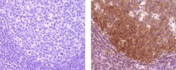
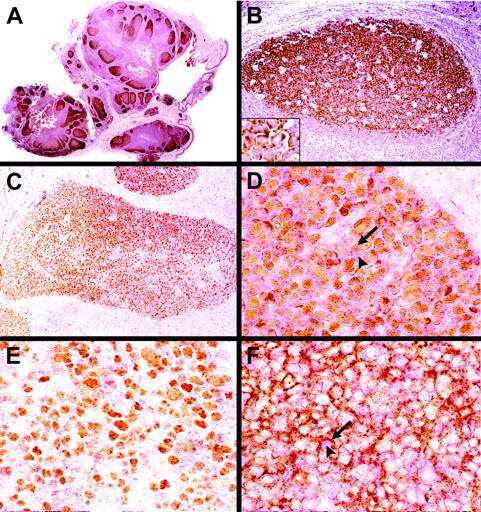
- Immunohistochemistry of formalin-fixed paraffin embedded human tonsil using 5 µg/mL of Mouse IgG2a K Isotype Control Purified (left) or 5 µg/mL of Anti-Human HGAL Purified (right) followed by Anti-Mouse IgG Biotin, Streptavidin HRP, and DAB visualization.Nuclei are counterstained with hematoxylin.

Supportive validation
- Submitted by
- Invitrogen Antibodies (provider)
- Main image

- Experimental details
- NULL
- Submitted by
- Invitrogen Antibodies (provider)
- Main image
- Experimental details
- NULL
- Submitted by
- Invitrogen Antibodies (provider)
- Main image
- Experimental details
- NULL

- Submitted by
- Invitrogen Antibodies (provider)
- Main image
- Experimental details
- NULL
- Submitted by
- Invitrogen Antibodies (provider)
- Main image
- Experimental details
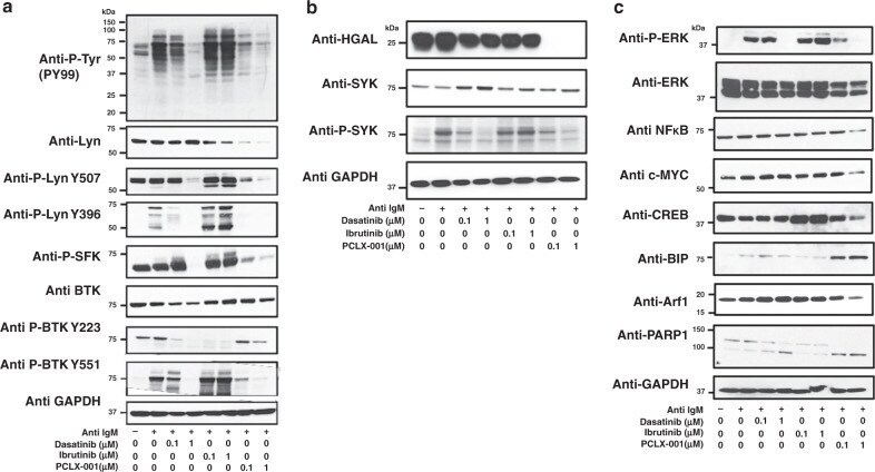
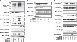
- Fig. 4 PCLX-001 treatment attenuates BCR downstream signaling events in BL2 lymphoma cells. Western blot of BL2 cells treated for 48 h with 0.1 uM or 1.0 muM of dasatinib, ibrutinib or PCLX-001 to detect total tyrosine phosphorylation (P-Tyr), Lyn, Lyn phosphorylated on tyrosine 396 or 507, BTK, and BTK phosphorylated on tyrosines 223 or 551 ( a ), HGAL, SYK, phosphorylated SYK (P-SYK) ( b ) or ERK, phosphorylated ERK (P-ERK), NFkappaB, c-Myc, CREB, Arf-1, BIP, and PARP-1 ( c ). Western blots are representative of at least three independent experiments. GAPDH serves as a loading control. BL2 cells were activated with 25 mug/mL F(ab') 2 anti-human IgM for 2 min and processed for western blotting. All western blots shown are representative of three independent experiments. Source data are provided as a Source Data file.