Explore
 Validate
 Learn
Learn Immunocytochemistry
Antibody data
- Antibody Data
- Antigen structure
- References [1]
- Comments [0]
- Validations
- Immunocytochemistry [1]
- Immunohistochemistry [2]
Submit
Validation data
Reference
Comment
Report error
- Product number
- MA5-16363 - Provider product page

- Provider
- Invitrogen Antibodies
- Product name
- Anti-F4/80 Monoclonal Antibody (SP115)
- Antibody type
- Monoclonal
- Antigen
- Synthetic peptide
- Description
- Heat-mediated antigen retrieval is recommended prior to staining, using a 10mM citrate buffer, pH 6.0, for 10 minutes followed by cooling at room temperature for 20 min. Following antigen retrieval, incubate samples with primary antibody for 10 min at room temperature. A suggested positive control is prostate tissue.
- Reactivity
- Human, Mouse
- Host
- Rabbit
- Isotype
- IgG
- Antibody clone number
- SP115
- Vial size
- 500 µL
- Concentration
- Lot Dependent
- Storage
- 4° C, do not freeze
Submitted references Glutathione S-transferase alpha 4 induction by activator protein 1 in colorectal cancer.
Yang Y, Huycke MM, Herman TS, Wang X
Oncogene 2016 Nov 3;35(44):5795-5806
Oncogene 2016 Nov 3;35(44):5795-5806
No comments: Submit comment
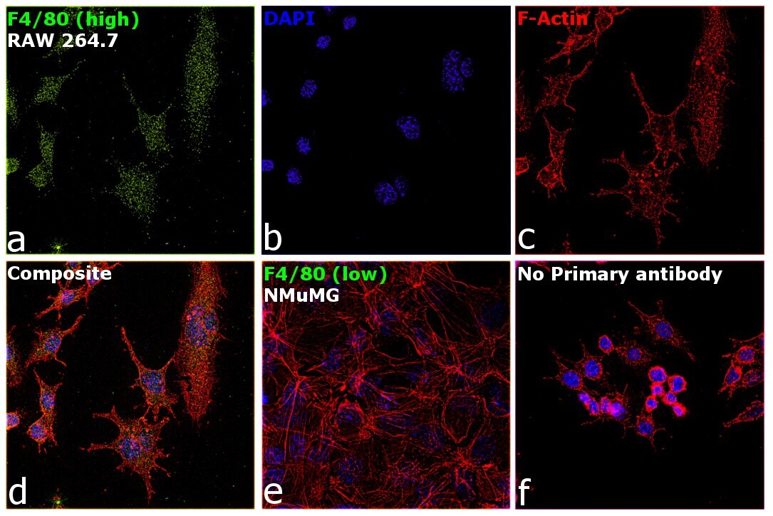
Supportive validation
- Submitted by
- Invitrogen Antibodies (provider)
- Main image

- Experimental details
- Immunofluorescence analysis of F4/80 was performed using 70% confluent log phase RAW 264.7 and NMuMG cells. The cells were fixed with 4% paraformaldehyde for 10 minutes, permeabilized with 0.1% Triton™ X-100 for 15 minutes, and blocked with 2% BSA for 1 hour at room temperature. The cells were labeled with F4/80 Monoclonal Antibody (CI:A3-1) (Product # MA5-16363) at 1:100 dilution in 0.1% BSA, incubated at 4 degree celsius overnight and then with Donkey anti-Rabbit IgG (H+L) Highly Cross-Adsorbed Secondary Antibody, Alexa Fluor Plus 488 (Product # A32790) at a dilution of 1:2000 for 45 minutes at room temperature (Panel a: green). Nuclei (Panel b: blue) were stained with SlowFade® Gold Antifade Mountant with DAPI (Product # S36938). F-actin (Panel c: red) was stained with Rhodamine Phalloidin (Product # R415, 1:300). Panel d represents the merged image showing cytoplasmic and membrane localization. Panel e represents NMuMG cells having no expression of F4/80. Panel f represents control cells with no primary antibody to assess background. The images were captured at 60X magnification.
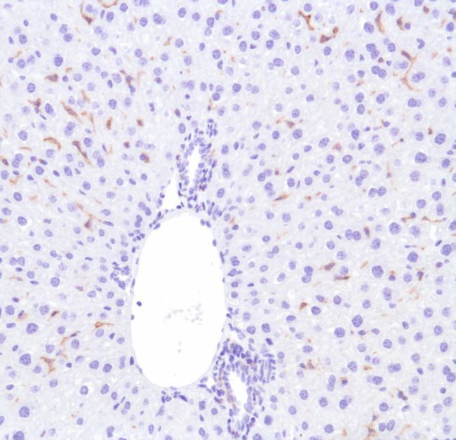
Supportive validation
- Submitted by
- Invitrogen Antibodies (provider)
- Main image

- Experimental details
- Immunohistochemical (paraffin) analysis of F4/80 using anti-F4/80 Monoclonal Antibody (Product # MA5-16363) in Mouse Liver Cancer Tissue. The recommended dilution for this antibody in immunohistochemistry applications is 1:100.
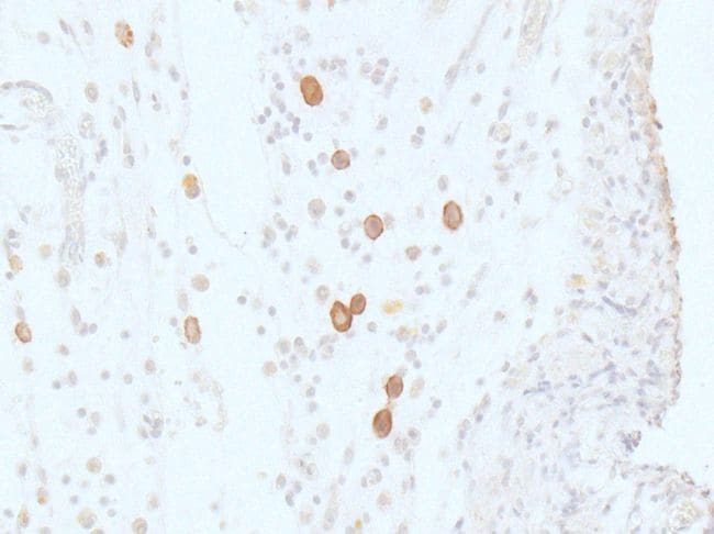
- Submitted by
- Invitrogen Antibodies (provider)
- Main image

- Experimental details
- Immunohistochemistry was performed on formalin -fixed paraffin-embedded rat infarct heart tissue sections. To expose target proteins, heat-induced epitope retrieval (HIER) was performed in sodium citrate buffer (pH 6.0) for 30 minutes at 100°C. Tissues were blocked in 10% normal goat serum for 20 minutes at room temperature and probed with a F4/80 monoclonal antibody (Product # MA5-16363) at a dilution of 1:100 for 1 hour at room temperature. Tissues were washed extensively with 1X PBS. Detection was performed using a biotinylated goat anti-rabbit IgG secondary antibody followed by an avidin-biotin complex reagent and DAB colorimetric substrate. Tissues were counterstained with hematoxilyn and visualized by light microscopy. Data courtesy of the Innovators Program.