Explore
 Validate
 Learn
Learn Western blot
 Immunocytochemistry
Antibody data
- Antibody Data
- Antigen structure
- References [3]
- Comments [0]
- Validations
- Immunocytochemistry [3]
- Immunohistochemistry [1]
- Other assay [4]
Submit
Validation data
Reference
Comment
Report error
- Product number
- PA5-21759 - Provider product page

- Provider
- Invitrogen Antibodies
- Product name
- AKAP12 Polyclonal Antibody
- Antibody type
- Polyclonal
- Antigen
- Synthetic peptide
- Description
- Recommended positive controls: 293T, A431, H1299, HeLaS3, HepG2. Predicted reactivity: Rhesus Monkey (100%). Store product as a concentrated solution. Centrifuge briefly prior to opening the vial.
- Reactivity
- Human
- Host
- Rabbit
- Isotype
- IgG
- Vial size
- 100 μL
- Concentration
- 0.21 mg/mL
- Storage
- Store at 4°C short term. For long term storage, store at -20°C, avoiding freeze/thaw cycles.
Submitted references Protein kinase A-mediated phosphorylation of naked cuticle homolog 2 stimulates cell-surface delivery of transforming growth factor-α for epidermal growth factor receptor transactivation.
The melanocortin signaling cAMP axis accelerates repair and reduces mutagenesis of platinum-induced DNA damage.
AKAP12 mediates PKA-induced phosphorylation of ATR to enhance nucleotide excision repair.
Cao Z, Singh B, Li C, Markham NO, Carrington LJ, Franklin JL, Graves-Deal R, Kennedy EJ, Goldenring JR, Coffey RJ
Traffic (Copenhagen, Denmark) 2019 May;20(5):357-368
Traffic (Copenhagen, Denmark) 2019 May;20(5):357-368
The melanocortin signaling cAMP axis accelerates repair and reduces mutagenesis of platinum-induced DNA damage.
Jarrett SG, Carter KM, Shelton BJ, D'Orazio JA
Scientific reports 2017 Sep 15;7(1):11708
Scientific reports 2017 Sep 15;7(1):11708
AKAP12 mediates PKA-induced phosphorylation of ATR to enhance nucleotide excision repair.
Jarrett SG, Wolf Horrell EM, D'Orazio JA
Nucleic acids research 2016 Dec 15;44(22):10711-10726
Nucleic acids research 2016 Dec 15;44(22):10711-10726
No comments: Submit comment
Supportive validation
- Submitted by
- Invitrogen Antibodies (provider)
- Main image
- Experimental details
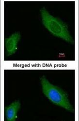
- Immunofluorescent analysis of AKAP12 in paraformaldehyde-fixed HeLa cells using an AKAP12 polyclonal antibody (Product # PA5-21759) at a 1:200 dilution.
- Submitted by
- Invitrogen Antibodies (provider)
- Main image
- Experimental details
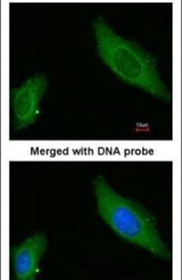
- Immunofluorescence analysis of paraformaldehyde-fixed HeLa, using AKAP12 (Product # PA5-21759) antibody at 1:200 dilution.

- Submitted by
- Invitrogen Antibodies (provider)
- Main image

- Experimental details
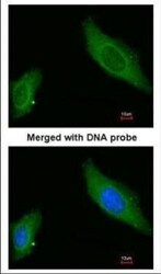
- Immunofluorescence analysis of paraformaldehyde-fixed HeLa, using AKAP12 (Product # PA5-21759) antibody at 1:200 dilution.

Supportive validation
- Submitted by
- Invitrogen Antibodies (provider)
- Main image
- Experimental details
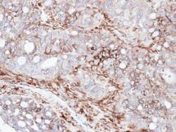
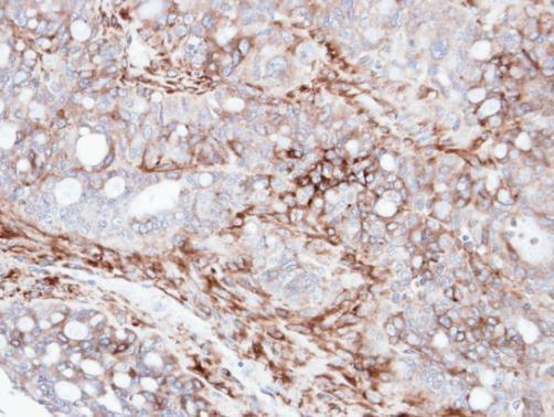
- Immunohistochemical analysis of paraffin-embedded human serous ovarian cancer, using AKAP12 (Product # PA5-21759) antibody at 1:100 dilution. Antigen Retrieval: EDTA based buffer, pH 8.0, 15 min.
Supportive validation
- Submitted by
- Invitrogen Antibodies (provider)
- Main image
- Experimental details
- NULL
- Submitted by
- Invitrogen Antibodies (provider)
- Main image
- Experimental details
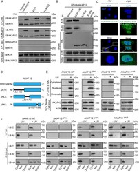
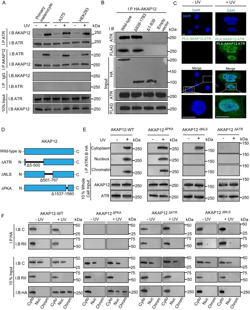
- Figure 1. AKAP12 interacts with ATR. ( A ) Cells were either mock-treated or exposed to UVB (10 J/m 2 ). After 30 min, whole cell lysates were sampled and reciprocal Co-IP and immunoblots of ATR and AKAP12 were performed. Input represents 10% of total cellular lysate. ( B ) Full-length HA-tagged AKAP12 and truncated HA-tagged AKAP12 mutants were transfected in HEK293 cells expressing FLAG-tagged ATR and exposed to UVB (10 J/m 2 ). Co-IP with anti-HA followed by immunoblotting with either anti-ATR or anti-FLAG at 30 min post-UVB. Input represents 10% of total cellular lysate. ( C ) Proximity ligation confirming sub-cellular localization of the ATR-AKAP12 interaction in HEK293 cells at 30 min after UVB (10 J/m 2 ) or mock treatment. PLA was performed with anti-AKAP12 and anti-ATR antibodies. Green detection events signify juxtaposition between AKAP12 and ATR in maximum intensity projection images. Nuclei were stained with DAPI (blue). Bar represents 50 mum. ( D ) Schematic diagram of wild-type AKAP12 and mutant variants that were engineered to either prevent the AKAP12-ATR interaction (AKAP12 DeltaATR ), prevent nuclear localization (AKAP12 DeltaNLS ) or prevent PKA-RII-AKAP12 binding (AKAP12 DeltaPKA ). ( E ) Co-IP of ATR and immunoblotting of HA-tagged wild-type or HA-tagged mutant AKAP12 (AKAP12 DeltaPKA , AKAP12 DeltaATR and AKAP12 DeltaNLS ). Cells were either mock-treated or exposed to UVB (10 J/m 2 ) and cytoplasm, nucleus and chromatin fractions extracted (30 min). Inpu
- Submitted by
- Invitrogen Antibodies (provider)
- Main image
- Experimental details
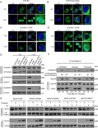
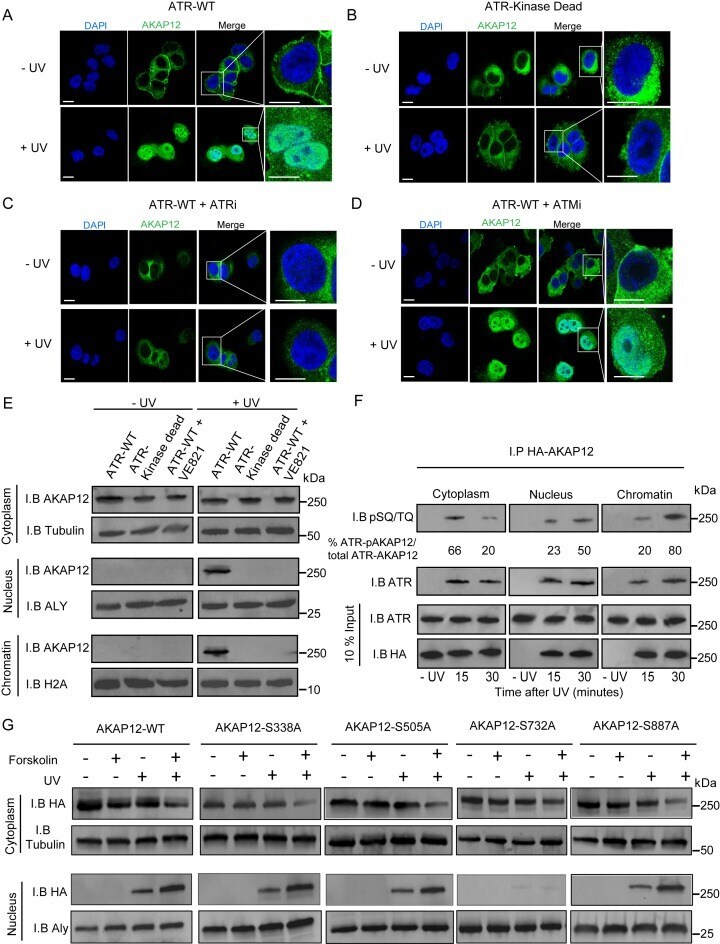
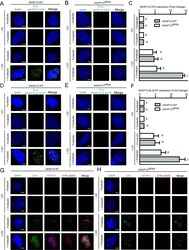
- Figure 2. ATR phosphorylates AKAP12. Confocal imaging of AKAP12 in HEK293 treated with siRNA-targeted to ATR and transfected with siRNA-resistant ( A ) wild-type ATR (ATR-WT) or ( B ) kinase-dead ATR (ATR-KD). Confocal imaging of AKAP12 in HEK293 cells expressing ATR-WT and treated with either an inhibitor against ( C ) ATR (VE-821; 10 muM) or ( D ) ATM (KU-55933; 10 muM). Cells were either mock-treated or exposed to UVB (10 J/m 2 ) and AKAP12 staining was determined 30 min post-damage. Green staining signifies AKAP12 maximum intensity projection images. Nuclei were stained with DAPI (blue). Bar represents 50 mum. ( E ) AKAP12 levels in cell fractions of ATR-siRNA silenced HEK293 cells transfected with siRNA-resistant wild-type (ATR-WT) or kinase-dead ATR (ATR-KD) and ATR-WT expressing HEK293 cells treated with ATR kinase inhibitor (VE-821; 10 muM). Cells were either mock-treated or exposed to UVB (10 J/m 2 ) and cytoplasmic, nuclear and chromatin fractions extracted. Input represents 10% of total cellular fraction. ( F ) Immunoprecipitation of AKAP12 in HEK293 cells expressing HA-tagged wild-type AKAP12 with an anti-HA antibody and immunoblotting with an ATR/ATM phosphorylation-specific antibody that detects phosphorylated SQ/TQ motifs in ATR-WT-transfected HEK293 cells at 15 and 30 min post-UVB (10 J/m 2 ). Percentages of ATR-phosphorylated-AKAP12 (ATR-pAKAP12) interactions from total ATR-AKAP12 interactions were determined from three experiments. Input represents 10% of re
- Submitted by
- Invitrogen Antibodies (provider)
- Main image
- Experimental details
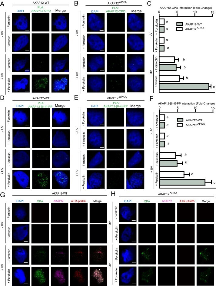
- Figure 6. AKAP12 interacts with UV-induced photoproducts. ( A and B ) AKAP12 CRISPR-deleted HEK293 cells were transfected with wild-type AKAP12 or AKAP12 DeltaPKA were treated with either vehicle or forskolin (10 muM) and exposed to UVC (10 J/m 2 ). The interaction between AKAP12 and CPDs at 30 min was determined by proximity ligation using anti-AKAP12 and anti-CPD antibodies. Green detection events signify juxtaposition between AKAP12 and [6-4]-PP in maximum intensity projection images. Nuclei were stained with DAPI (blue). Bar represents 50 mum. ( C ) Quantification of the AKAP12-CPD localization shown in panel A and B. At least 100 cells were counted from representative fields from two separate experiments. Values not sharing a common letter were significantly different as determined by one-way ANOVA; P