 Explore
 Validate
 Learn
Learn Western blot
 Flow cytometry
Antibody data
- Antibody Data
- Antigen structure
- References [4]
- Comments [0]
- Validations
- Flow cytometry [1]
Submit
Validation data
Reference
Comment
Report error
- Product number
- 14-1159-82 - Provider product page

- Provider
- Invitrogen Antibodies
- Product name
- CD115 (c-fms) Monoclonal Antibody (12-3A3-1B10), eBioscience™
- Antibody type
- Monoclonal
- Antigen
- Other
- Description
- Description: CD115 or Colony-Stimulating Factor-1 Receptor (CSF-1R) is expressed on cells committed to the monocyte lineage and osteoclasts. CSF-1R is a disulfide-linked homodimer, which upon binding of its ligand, CSF-1, undergoes tyrosine autophosphorylation and subsequently, phosphorylates other membrane-proximal downstream targets which results in cytoskeletal remodeling, gene transcription and protein translation. CSF-1R activation promotes the survival, proliferation and differentiation of mononuclear phagocytes and the spreading and motility of macrophages. In osteoclasts, CSF-1R synergizes with RANKL to regulate the differentiation of mononuclear phagocytes to osteoclasts. Applications Reported: This 12-3A3-1B10 antibody has been reported for use in flow cytometric analysis, immunoprecipitation, immunoblotting (WB), and immunohistochemical staining. Applications Tested: This 12-3A3-1B10 antibody has been tested by flow cytometric analysis of normal human peripheral blood cells. This can be used at less than or equal to 1 µg per test. A test is defined as the amount (µg) of antibody that will stain a cell sample in a final volume of 100 µL. Cell number should be determined empirically but can range from 10^5 to 10^8 cells/test. It is recommended that the antibody be carefully titrated for optimal performance in the assay of interest. Purity: Greater than 90%, as determined by SDS-PAGE. Aggregation: Less than 10%, as determined by HPLC. Filtration: 0.2 µm post-manufacturing filtered.
- Reactivity
- Human
- Host
- Rat
- Isotype
- IgG
- Antibody clone number
- 12-3A3-1B10
- Vial size
- 100 μg
- Concentration
- 0.5 mg/mL
- Storage
- 4°C
Submitted references A unique anti-CD115 monoclonal antibody which inhibits osteolysis and skews human monocyte differentiation from M2-polarized macrophages toward dendritic cells.
Comparison of gene expression profiles between human and mouse monocyte subsets.
CSF-1 regulation of the wandering macrophage: complexity in action.
Monoclonal antibodies to the human CSF-1 receptor (c-fms proto-oncogene product) detect epitopes on normal mononuclear phagocytes and on human myeloid leukemic blast cells.
Haegel H, Thioudellet C, Hallet R, Geist M, Menguy T, Le Pogam F, Marchand JB, Toh ML, Duong V, Calcei A, Settelen N, Preville X, Hennequi M, Grellier B, Ancian P, Rissanen J, Clayette P, Guillen C, Rooke R, Bonnefoy JY
mAbs 2013 Sep-Oct;5(5):736-47
mAbs 2013 Sep-Oct;5(5):736-47
Comparison of gene expression profiles between human and mouse monocyte subsets.
Ingersoll MA, Spanbroek R, Lottaz C, Gautier EL, Frankenberger M, Hoffmann R, Lang R, Haniffa M, Collin M, Tacke F, Habenicht AJ, Ziegler-Heitbrock L, Randolph GJ
Blood 2010 Jan 21;115(3):e10-9
Blood 2010 Jan 21;115(3):e10-9
CSF-1 regulation of the wandering macrophage: complexity in action.
Pixley FJ, Stanley ER
Trends in cell biology 2004 Nov;14(11):628-38
Trends in cell biology 2004 Nov;14(11):628-38
Monoclonal antibodies to the human CSF-1 receptor (c-fms proto-oncogene product) detect epitopes on normal mononuclear phagocytes and on human myeloid leukemic blast cells.
Ashmun RA, Look AT, Roberts WM, Roussel MF, Seremetis S, Ohtsuka M, Sherr CJ
Blood 1989 Feb 15;73(3):827-37
Blood 1989 Feb 15;73(3):827-37
No comments: Submit comment
Supportive validation
- Submitted by
- Invitrogen Antibodies (provider)
- Main image
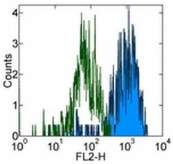
- Experimental details
- Staining of normal human peripheral blood cells with 0.5 µg of Rat IgG2a K Isotype Control Purified (Product # 14-4321-82) (open histogram) or 0.5 µg of Anti-Human CD115 (c-fms) Purified (filled histogram) followed by Anti-Rat IgG Biotin (Product # 13-4813-85) and Streptavidin PE (Product # 12-4317-87). Cells in the monocyte gate were used for analysis.
