Explore
 Validate
 Learn
Learn Western blot
 Immunocytochemistry
Antibody data
- Antibody Data
- Antigen structure
- References [2]
- Comments [0]
- Validations
- Immunocytochemistry [4]
- Flow cytometry [2]
- Other assay [1]
Submit
Validation data
Reference
Comment
Report error
- Product number
- PA5-25974 - Provider product page

- Provider
- Invitrogen Antibodies
- Product name
- CSF1R Polyclonal Antibody
- Antibody type
- Polyclonal
- Antigen
- Recombinant full-length protein
- Reactivity
- Human, Mouse
- Host
- Rabbit
- Isotype
- IgG
- Vial size
- 400 μL
- Concentration
- 2 mg/mL
- Storage
- Store at 4°C short term. For long term storage, store at -20°C, avoiding freeze/thaw cycles.
Submitted references Minocycline suppresses disease-associated microglia (DAM) in a model of photoreceptor cell degeneration.
Evidence of a Myenteric Plexus Barrier and Its Macrophage-Dependent Degradation During Murine Colitis: Implications in Enteric Neuroinflammation.
Ozaki E, Delaney C, Campbell M, Doyle SL
Experimental eye research 2022 Apr;217:108953
Experimental eye research 2022 Apr;217:108953
Evidence of a Myenteric Plexus Barrier and Its Macrophage-Dependent Degradation During Murine Colitis: Implications in Enteric Neuroinflammation.
Dora D, Ferenczi S, Stavely R, Toth VE, Varga ZV, Kovacs T, Bodi I, Hotta R, Kovacs KJ, Goldstein AM, Nagy N
Cellular and molecular gastroenterology and hepatology 2021;12(5):1617-1641
Cellular and molecular gastroenterology and hepatology 2021;12(5):1617-1641
No comments: Submit comment
Supportive validation
- Submitted by
- Invitrogen Antibodies (provider)
- Main image
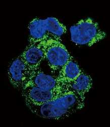
- Experimental details
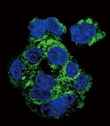
- Immunofluorescent analysis of HepG2 cells using a CSF1R polyclonal antibody (Product # PA5-25974) at a dilution of 1:10-50, followed by a fluor-conjugated goat anti-rabbit secondary antibody (green). Nuclei were stained with DAPI (blue).
- Submitted by
- Invitrogen Antibodies (provider)
- Main image

- Experimental details
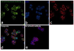
- Immunofluorescence analysis of CSF1R was performed using 70% confluent log phase THP-1 cells. The cells were fixed with 4% paraformaldehyde for 10 minutes, permeabilized with 0.1% Triton™ X-100 for 15 minutes, and blocked with 1% BSA for 1 hour at room temperature. The cells were labeled with anti-CSF1R Polyclonal Antibody (Product # PA5-25974) at 1:200 dilution in 0.1% BSA, incubated at 4 degree Celsius overnight and then labeled with Goat anti-Rabbit IgG (H+L) Superclonal™ Secondary Antibody, Alexa Fluor® 488 conjugate (Product # A27034) at a dilution of 1:2000 for 45 minutes at room temperature (Panel a: green). Nuclei (Panel b: blue) were stained with ProLong™ Diamond Antifade Mountant with DAPI (Product # P36962). F-actin (Panel c: red) was stained with Rhodamine Phalloidin (Product # R415, 1:300). Panel d represents the merged image showing Plasma Membrane localization. Panel e represents control cells with no primary antibody to assess background. The images were captured at 60X magnification
- Submitted by
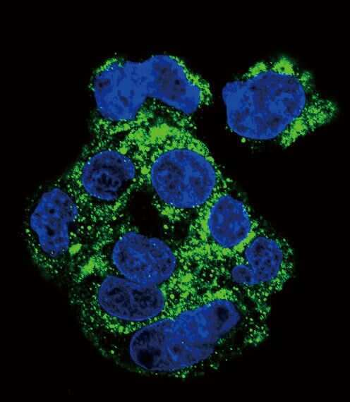
- Invitrogen Antibodies (provider)
- Main image
- Experimental details
- Immunocytochemistry analysis of CSF1R in HepG2 cells. Samples were incubated in CSF1R polyclonal antibody (Product # PA5-25974) followed by Alexa Fluor 488-conjugated goat anti-rabbit lgG (green). DAPI was used to stain the cell nuclear (blue).

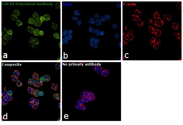
- Submitted by
- Invitrogen Antibodies (provider)
- Main image
- Experimental details
- Immunofluorescence analysis of CSF1R was performed using 70% confluent log phase THP-1 cells. The cells were fixed with 4% paraformaldehyde for 10 minutes, permeabilized with 0.1% Triton™ X-100 for 15 minutes, and blocked with 1% BSA for 1 hour at room temperature. The cells were labeled with anti-CSF1R Polyclonal Antibody (Product # PA5-25974) at 1:200 dilution in 0.1% BSA, incubated at 4 degree Celsius overnight and then labeled with Goat anti-Rabbit IgG (Heavy Chain) Superclonal™ Secondary Antibody, Alexa Fluor® 488 conjugate (Product # A27034) at a dilution of 1:2000 for 45 minutes at room temperature (Panel a: green). Nuclei (Panel b: blue) were stained with ProLong™ Diamond Antifade Mountant with DAPI (Product # P36962). F-actin (Panel c: red) was stained with Rhodamine Phalloidin (Product # R415, 1:300). Panel d represents the merged image showing Plasma Membrane localization. Panel e represents control cells with no primary antibody to assess background. The images were captured at 60X magnification

Supportive validation
- Submitted by
- Invitrogen Antibodies (provider)
- Main image

- Experimental details
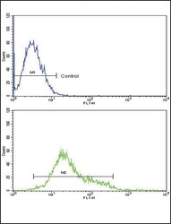
- Flow cytometry analysis of K562 cells using a CD115/MCSF Receptor polyclonal antibody (Product # PA5-25974) (bottom) compared to a negative control cell (top) at a dilution of 1:10-50, followed by a FITC-conjugated goat anti-rabbit antibody
- Submitted by
- Invitrogen Antibodies (provider)
- Main image
- Experimental details
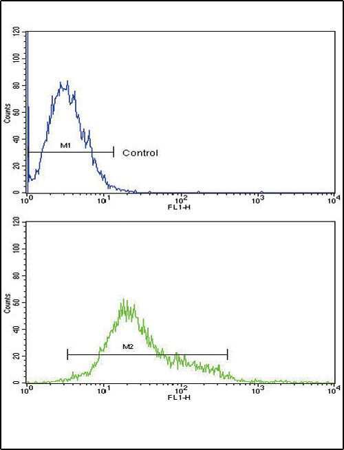
- Flow cytometry of CSF1R in K562 cells (bottom histogram). Samples were incubated with CSF1R polyclonal antibody (Product # PA5-25974) followed by FITC-conjugated goat-anti-rabbit secondary antibody. Negative control (top histogram).

Supportive validation
- Submitted by
- Invitrogen Antibodies (provider)
- Main image
- Experimental details
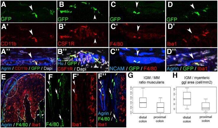
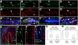
- Figure 2 Immunophenotype and spatial distribution of IGMs in CX3CR1 GFP mouse colon. GFP+ IGMs are present within the agrin-bordered enteric ganglia and co-express CD11b ( A-A"" ), CSF1R ( B-B"" ), F4/80 ( C-C"" ), and Iba1 ( D-D"" ). F4/80 and Iba1 are co-expressed by MMs and IGMs ( F-F"" , arrowheads , magnified from circled area in E ), but Iba1 is not present in submucosal macrophages ( F-F"" , arrows ). In lymphatic aggregates of the colonic mucosa ( E , area within dashed line ), macrophages express Iba1 but not F4/80. Comparison of MM and IGM cell number in the proximal and distal colon of CX3CR1 GFP mice shows no significant difference in ratio of IGMs to total MMs ( G ; 3.26 % +- 1.3 % vs 2.12 % +- 1.1 %, P = .0977, n = 8). However, the number of IGMs is significantly higher in distal colon compared with proximal colon when adjusted to myenteric ganglion area ( H ; 82.37 +- 32.98 vs 50.25 +- 19.6 cell/mm 2 , P = .0309, n = 8).