Explore
 Validate
 Learn
Learn Western blot
 Immunocytochemistry
Antibody data
- Antibody Data
- Antigen structure
- References [4]
- Comments [0]
- Validations
- Immunocytochemistry [1]
Submit
Validation data
Reference
Comment
Report error
- Product number
- HPA012323 - Provider product page

- Provider
- Atlas Antibodies
- Proper citation
- Atlas Antibodies Cat#HPA012323, RRID:AB_1848977
- Product name
- Anti-CSF1R
- Antibody type
- Polyclonal
- Description
- Polyclonal Antibody against Human CSF1R, Gene description: colony stimulating factor 1 receptor, Alternative Gene Names: C-FMS, CD115, CSFR, FMS, Validated applications: WB, IHC, ICC, Uniprot ID: P07333, Storage: Store at +4°C for short term storage. Long time storage is recommended at -20°C.
- Reactivity
- Human
- Host
- Rabbit
- Conjugate
- Unconjugated
- Isotype
- IgG
- Vial size
- 100 µl
- Concentration
- 0.1 mg/ml
- Storage
- Store at +4°C for short term storage. Long time storage is recommended at -20°C.
- Handling
- The antibody solution should be gently mixed before use.
Submitted references High Expression of CSF-1R Predicts Poor Prognosis and CSF-1Rhigh Tumor-Associated Macrophages Inhibit Anti-Tumor Immunity in Colon Adenocarcinoma
Characterization of a p53/miR-34a/CSF1R/STAT3 Feedback Loop in Colorectal Cancer
High co-expression of IL-34 and M-CSF correlates with tumor progression and poor survival in lung cancers
Inhibition of the colony-stimulating-factor-1 receptor affects the resistance of lung cancer cells to cisplatin
Wang X, Zhang J, Hu B, Qian F
Frontiers in Oncology 2022;12
Frontiers in Oncology 2022;12
Characterization of a p53/miR-34a/CSF1R/STAT3 Feedback Loop in Colorectal Cancer
Shi X, Kaller M, Rokavec M, Kirchner T, Horst D, Hermeking H
Cellular and Molecular Gastroenterology and Hepatology 2020;10(2):391-418
Cellular and Molecular Gastroenterology and Hepatology 2020;10(2):391-418
High co-expression of IL-34 and M-CSF correlates with tumor progression and poor survival in lung cancers
Baghdadi M, Endo H, Takano A, Ishikawa K, Kameda Y, Wada H, Miyagi Y, Yokose T, Ito H, Nakayama H, Daigo Y, Suzuki N, Seino K
Scientific Reports 2018;8(1)
Scientific Reports 2018;8(1)
Inhibition of the colony-stimulating-factor-1 receptor affects the resistance of lung cancer cells to cisplatin
Pass H, Lavilla C, Canino C, Goparaju C, Preiss J, Noreen S, Blandino G, Cioce M
Oncotarget 2016;7(35):56408-56421
Oncotarget 2016;7(35):56408-56421
No comments: Submit comment
Supportive validation
- Submitted by
- Atlas Antibodies (provider)
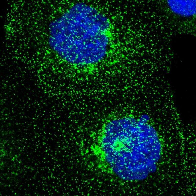
- Main image
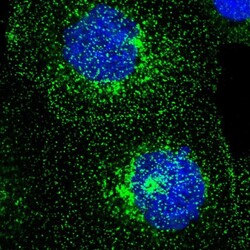
- Experimental details
- Immunofluorescent staining of human cell line A-431 shows localization to plasma membrane & vesicles.
- Sample type
- Human