Explore
 Validate
 Learn
Learn Western blot
 Immunocytochemistry
Antibody data
- Antibody Data
- Antigen structure
- References [2]
- Comments [0]
- Validations
- Western blot [1]
- Immunocytochemistry [1]
- Immunohistochemistry [1]
Submit
Validation data
Reference
Comment
Report error
- Product number
- HPA023505 - Provider product page

- Provider
- Atlas Antibodies
- Proper citation
- Atlas Antibodies Cat#HPA023505, RRID:AB_1845148
- Product name
- Anti-ATF7IP
- Antibody type
- Polyclonal
- Description
- Polyclonal Antibody against Human ATF7IP, Gene description: activating transcription factor 7 interacting protein, Alternative Gene Names: FLJ10688, p621, Validated applications: IHC, ICC, Uniprot ID: Q6VMQ6, Storage: Store at +4°C for short term storage. Long time storage is recommended at -20°C.
- Reactivity
- Human
- Host
- Rabbit
- Conjugate
- Unconjugated
- Isotype
- IgG
- Vial size
- 100 µl
- Concentration
- 0.3 mg/ml
- Storage
- Store at +4°C for short term storage. Long time storage is recommended at -20°C.
- Handling
- The antibody solution should be gently mixed before use.
Submitted references CRISPR screens identify gene targets at breast cancer risk loci
Conserved transcription factors promote cell fate stability and restrict reprogramming potential in differentiated cells
Tuano N, Beesley J, Manning M, Shi W, Perlaza-Jimenez L, Malaver-Ortega L, Paynter J, Black D, Civitarese A, McCue K, Hatzipantelis A, Hillman K, Kaufmann S, Sivakumaran H, Polo J, Reddel R, Band V, French J, Edwards S, Powell D, Chenevix-Trench G, Rosenbluh J
Genome Biology 2023;24(1)
Genome Biology 2023;24(1)
Conserved transcription factors promote cell fate stability and restrict reprogramming potential in differentiated cells
Missinato M, Murphy S, Lynott M, Yu M, Kervadec A, Chang Y, Kannan S, Loreti M, Lee C, Amatya P, Tanaka H, Huang C, Puri P, Kwon C, Adams P, Qian L, Sacco A, Andersen P, Colas A
Nature Communications 2023;14(1)
Nature Communications 2023;14(1)
No comments: Submit comment
Enhanced validation
- Submitted by
- klas2
- Enhanced method
- Genetic validation
- Main image
- Experimental details
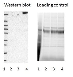
- Western blot of cell lysate from U-2 OS cells transfected with either siRNA targeting ATF7IP or control siRNA. Lane 1: Marker (250, 130, 95, 72, 55, 36, 28, 17, 10) Lane 2: Cell lysate from U-2OS cells transfected with siRNA targeting ATF7IP Lane 3: N/A Lane 4: Cell lysate from U-2OS cells transfected with control siRNA Right image, lane 1-4: loading control
- Sample type
- U-2 OS
- Primary Ab dilution
- 1:383
- Conjugate
- Horseradish Peroxidase
- Secondary Ab
- Secondary Ab
- Secondary Ab dilution
- 1:3000
- Knockdown/Genetic Approaches Application
- Western blot
Supportive validation
- Submitted by
- Atlas Antibodies (provider)
- Main image

- Experimental details
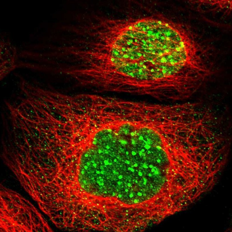
- Immunofluorescent staining of human cell line A-431 shows localization to nucleoplasm & nuclear bodies.
- Sample type
- Human
Supportive validation
- Submitted by
- Atlas Antibodies (provider)
- Enhanced method
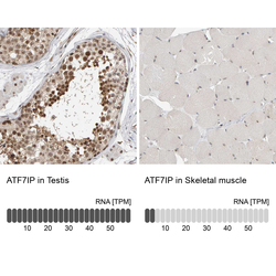
- Orthogonal validation
- Main image
- Experimental details
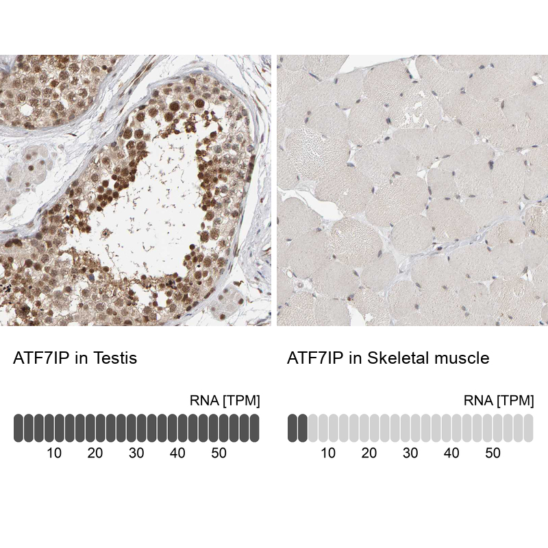
- Immunohistochemistry analysis in human testis and skeletal muscle tissues using HPA023505 antibody. Corresponding ATF7IP RNA-seq data are presented for the same tissues.
- Sample type
- Human
- Protocol
- Protocol