Explore
 Validate
 Learn
Learn Western blot
Antibody data
- Antibody Data
- Antigen structure
- References [0]
- Comments [0]
- Validations
- Western blot [5]
- Immunocytochemistry [2]
- Immunohistochemistry [1]
- Flow cytometry [1]
- Other assay [6]
Submit
Validation data
Reference
Comment
Report error
- Product number
- 36-8800 - Provider product page

- Provider
- Invitrogen Antibodies
- Product name
- Phospho-ERK1/ERK2 (Thr202, Tyr204) Polyclonal Antibody
- Antibody type
- Polyclonal
- Antigen
- Synthetic peptide
- Description
- This antibody is predicted to react with Xenopus and zebrafish based on 100% sequence homology. 36-8800 is specific for the ~42k - 44k ERK/MAPK phosphorylated at Thr202 and Tyr204. Immunolabeling is blocked by the phosphopeptide used as antigen but not by the corresponding dephosphopeptide. The immunolabeling is completely eliminated by lambda-phosphatase.
- Reactivity
- Human, Mouse, Rat
- Host
- Rabbit
- Isotype
- IgG
- Vial size
- 100 µL
- Storage
- -20° C, Avoid Freeze/Thaw Cycles
No comments: Submit comment
Supportive validation
- Submitted by
- Invitrogen Antibodies (provider)
- Main image

- Experimental details
- Western blot analysis of Phospho-MAPK1/3 pThr202/Tyr204 was performed by loading rat brain lysates onto a SDS-PAGE gel. Proteins were transferred to a membrane, blocked, and probed with a Phospho-MAPK1/3 pThr202/Tyr204 polyclonal antibody (Product # 36-8800) at a dilution of 1:1000, followed by an HRP-conjugated anti-rabbit IgG secondary antibody. Detection was performed using a chemiluminescent substrate.

- Submitted by
- Invitrogen Antibodies (provider)
- Main image
- Experimental details
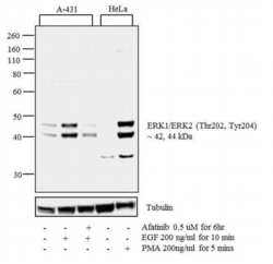
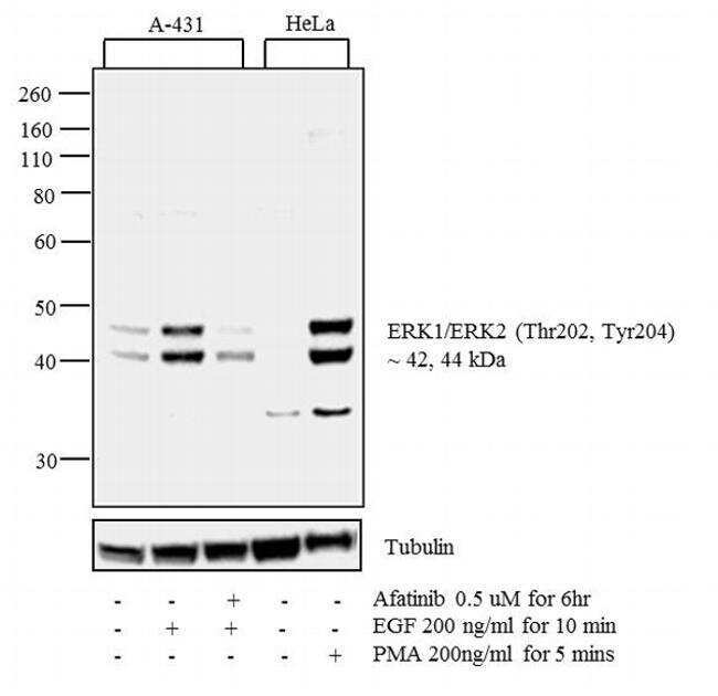
- Western blot analysis was performed on whole cell extracts (30 µg lysate) of A-431 (1), A-431 treated with EGF (200 ng/mL for 10 minutes) (2), A-431 treated with Afatinib followed by EGF (0.5 uM for 6 hours, 200 ng/mL for 10 minutes) (3), HeLa (4) and HeLa treated with PMA (200 ng/mL for 5 min). The blot was probed with Anti-Phospho-ERK1/ERK2 (Thr202, Tyr204) Rabbit Polyclonal Antibody (Product # 36-8800, 1:250 dilution) and detected by chemiluminescence using Goat anti-Rabbit IgG (H+L) Superclonal™ Secondary Antibody, HRP conjugate (Product # A27036, 0.25 µg/mL, 1:4000 dilution). 42, 44 kDa band corresponding to Phospho-ERK1/ERK2 (Thr202, Tyr204) was detected to increase upon EGF and PMA treatments in the cell lines tested. Pre-treatment with EGFR-antagonist, Afatinib, resulted in inhibition of Phospho-ERK1/ERK2 (Thr202, Tyr204) in A-431 cell line. Known quantity of protein samples were electrophoresed using Novex® NuPAGE® 4-12 % Bis-Tris gel (Product # NP0321BOX), XCell SureLock System (Product # EI0002) and Novex Protein Standard (Product # LC5800). Proteins were then transferred onto a nitrocellulose membrane with iBlot® 2 Dry Blotting System (Product # IB21001). The membrane was probed with the relevant primary and secondary Antibody following blocking with 5 % skimmed milk. Chemiluminescent detection was performed using Pierce™ ECL Western Blotting Substrate (Product # 32106).
- Submitted by
- Invitrogen Antibodies (provider)
- Main image

- Experimental details
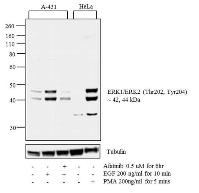
- Western blot analysis was performed on whole cell extracts (30 µg lysate) of A-431 (1), A-431 treated with EGF (200 ng/mL for 10 minutes) (2), A-431 treated with Afatinib followed by EGF (0.5 uM for 6 hours, 200 ng/mL for 10 minutes) (3), HeLa (4) and HeLa treated with PMA (200 ng/mL for 5 min). The blot was probed with Anti-Phospho-ERK1/ERK2 (Thr202, Tyr204) Rabbit Polyclonal Antibody (Product # 36-8800, 1:250 dilution) and detected by chemiluminescence using Goat anti-Rabbit IgG (H+L) Superclonal™ Secondary Antibody, HRP conjugate (Product # A27036, 0.25 µg/mL, 1:4000 dilution). 42, 44 kDa band corresponding to Phospho-ERK1/ERK2 (Thr202, Tyr204) was detected to increase upon EGF and PMA treatments in the cell lines tested. Pre-treatment with EGFR-antagonist, Afatinib, resulted in inhibition of Phospho-ERK1/ERK2 (Thr202, Tyr204) in A-431 cell line. Known quantity of protein samples were electrophoresed using Novex® NuPAGE® 4-12 % Bis-Tris gel (Product # NP0321BOX), XCell SureLock System (Product # EI0002) and Novex Protein Standard (Product # LC5800). Proteins were then transferred onto a nitrocellulose membrane with iBlot® 2 Dry Blotting System (Product # IB21001). The membrane was probed with the relevant primary and secondary Antibody following blocking with 5 % skimmed milk. Chemiluminescent detection was performed using Pierce™ ECL Western Blotting Substrate (Product # 32106).
- Submitted by
- Invitrogen Antibodies (provider)
- Main image
- Experimental details
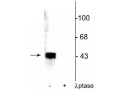
- Western blot of ERK1/2 in human T47D cell lysate showing specific immunolabeling of a ~42-44 kDa band corresponding to Phospho-ERK1/ERK2 (Thr202, Tyr204) polyclonal antibody (Product # 36-8800) in the first lane (-). Phosphospecificity is shown in the second lane (+) where immunolabeling is completely eliminated by blot treatment with lambda phosphatase (1,200 units for 30 min).

- Submitted by
- Invitrogen Antibodies (provider)
- Main image

- Experimental details
- Western blot analysis of p44 MAPK + p42 MAPK (pT202 + pT204) was performed by loading 30 µg of A431 (lane1), A431 treated for 10 minutes with 50 ng/mL PDGF (lane2), HeLa (lane3), HeLa treated for 10 minutes with 1:250 dilution of PDGF (lane4), NIH/3T3 (lane5), PC-12 (lane6), A549 (lane7), lysate using Novex® NuPAGE® 4-12 % Bis-Tris gel (Product # NP0322BOX), XCell SureLock™ Electrophoresis System (Product # EI0002), Novex® Sharp Pre-Stained Protein Standard (LC5800), and iBlot® Dry Blotting System (IB21001). Proteins were transferred to a nitrocellulose membrane and blocked with 5 % skim milk for 1 hour at room temperature. p44 MAPK + p42 MAPK (pT202 + pT204) was detected at 42, 44 kDa using p44 MAPK + p42 MAPK (pT202 + pT204) Rabbit Polyclonal Antibody (Product # 36-8800) at 1- 3 µg/mL in 5 % skim milk at 4°C overnight on a rocking platform. Goat Anti-Rabbit IgG - HRP Secondary Antibody (G21234) at 1:5000 dilution was used and chemiluminescent detection was performed using Pierce™ ECL Western Blotting Substrate (Product # 32106).

Supportive validation
- Submitted by
- Invitrogen Antibodies (provider)
- Main image

- Experimental details
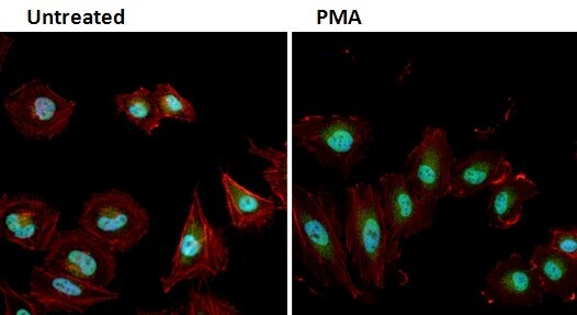
- Immunofluorescent analysis of Phospho-MAPK1/3 pThr202/Tyr204 (green) in HeLa cells either left untreated (left panel) or treated with 50nM PMA (right panel) for 10 minutes. Formalin fixed cells were permeabilized with 0.1% Triton X-100 in TBS for 10 minutes at room temperature and blocked with 1% Blocker BSA (Product # 37525) for 15 minutes at room temperature. Cells were probed with a Phospho-MAPK1/3 pThr202/Tyr204 polyclonal antibody (Product # 36-8800) at a dilution of 1:100 for at least 1 hour at room temperature, washed with PBS, and incubated with DyLight 488 goat anti-rabbit IgG secondary antibody (Product # 35552) at a dilution of 1:400 for 30 minutes at room temperature. F-Actin (red) was stained with DyLight 554 Phalloidin (Product # 21834) and nuclei (blue) were stained with Hoechst 33342 dye (Product # 62249). Images were taken on a Thermo Scientific ArrayScan or ToxInsight Instrument at 20X magnification.
- Submitted by
- Invitrogen Antibodies (provider)
- Main image
- Experimental details
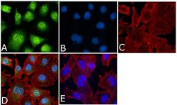
- Immunofluorescent analysis of Phospho-p44 MAPK + p42 MAPK pThr202 + pTyr204 Antibody was done on 70% confluent log phase A549 cells. The cells were fixed with 4% paraformaldehyde for 15 minutes, permeabilized with 0.25% Triton™ X-100 for 10 minutes, and blocked with 5% BSA for 1 hour at room temperature. The cells were labeled with Phospho-p44 MAPK + p42 MAPK pThr202 + pTyr204 Antibody (Product # 36-8800) at 1:250 dilution in 1% BSA and incubated for 3 hours at room temperature and then labeled with Alexa Fluor 488 Goat Anti-Rabbit IgG Secondary Antibody (Product # A-11008) at a dilution of 1:400 for 45 minutes at room temperature (Panel a: green). Nuclei (Panel b: blue) were stained with SlowFade® Gold Antifade Mountant with DAPI (Product # S36938). F-actin (Panel c: red) was stained with Alexa Fluor 594 Phalloidin (Product # A12381). Panel d is a merged image showing cytoplasmic and nuclear localization. Panel e is a no primary antibody control. The images were captured at 40X magnification.

Supportive validation
- Submitted by
- Invitrogen Antibodies (provider)
- Main image

- Experimental details
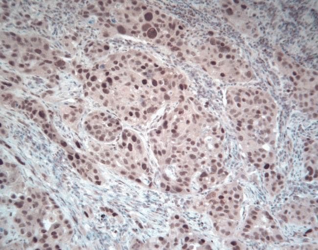
- Immunohistochemical staining of paraffin-embedded human lung cancer tissue using Zymed Rb anti-phospho-ERK1+2 (Thr202/Tyr204) (Product # 36-8800).
Supportive validation
- Submitted by
- Invitrogen Antibodies (provider)
- Main image

- Experimental details
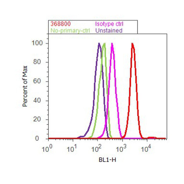
- Flow cytometry analysis of p44 MAPK+ p42 MAPK [pT202+ pY204] was done on K562 cells treated with TPA (200nM, 20 minutes). Cells were fixed with 70% ethanol for 10 minutes, permeabilized with 0.25% Triton™ X-100 for 20 minutes, and blocked with 5% BSA for 30 minutes at room temperature. Cells were labeled with p44 MAPK+ p42 MAPK [pT202+ pY204] Rabbit Polyclonal Antibody (368800, red histogram) or with rabbit isotype control (pink histogram) at 3-5 ug/million cells in 2.5% BSA. After incubation at room temperature for 2 hours, the cells were labeled with Alexa Fluor® 488 Goat Anti-Rabbit Secondary Antibody (A11008) at a dilution of 1:400 for 30 minutes at room temperature. The representative 10,000 cells were acquired and analyzed for each sample using an Attune® Acoustic Focusing Cytometer. The purple histogram represents unstained control cells and the green histogram represents no-primary-antibody control.
Supportive validation
- Submitted by
- Invitrogen Antibodies (provider)
- Main image

- Experimental details
- NULL
- Submitted by
- Invitrogen Antibodies (provider)
- Main image
- Experimental details
- NULL

- Submitted by
- Invitrogen Antibodies (provider)
- Main image

- Experimental details
- NULL
- Submitted by
- Invitrogen Antibodies (provider)
- Main image

- Experimental details
- NULL

- Submitted by
- Invitrogen Antibodies (provider)
- Main image
- Experimental details
- NULL

- Submitted by
- Invitrogen Antibodies (provider)
- Main image
- Experimental details
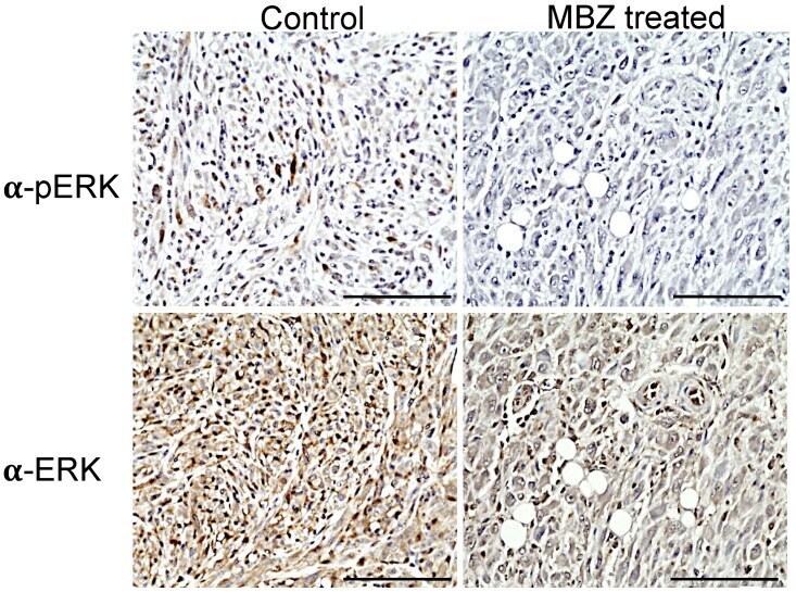
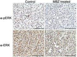
- Figure 4 MBZ reduces ERK (pERK) in treated NPcis mice. Representative images of tumors from untreated controls (left) and MBZ-treated NPcis mice (left) were stained for pERK1/2 (upper row) and ERK1/2 (lower row). pERK staining was visualized in brown in untreated controls but reduced in tumors of MBZ-treated mice. Each scale bar represents 100 um.