Explore
 Validate
 Learn
Learn Immunocytochemistry
 Immunohistochemistry
Antibody data
- Antibody Data
- Antigen structure
- References [0]
- Comments [0]
- Validations
- Immunocytochemistry [1]
- Other assay [4]
Submit
Validation data
Reference
Comment
Report error
- Product number
- 41-9108-82 - Provider product page

- Provider
- Invitrogen Antibodies
- Product name
- ERK1/2 Monoclonal Antibody (5AD13MA), eFluor™ 570, eBioscience™
- Antibody type
- Monoclonal
- Antigen
- Other
- Description
- Description: The 5AD13MA monoclonal antibody recognizes human, mouse, and rat extracellular signal-regulated kinases 1 and 2 (also known as ERK1/2, p44/42, or MAPK3/1). ERK1/2 belong to a family of conserved serine/threonine protein kinases known as mitogen-activated protein kinases (MAPKs) that are involved in many cellular programs such as proliferation, differentiation, motility, and survival. The ERK1/2 signaling pathway can be activated in response to a diverse range of extracellular stimuli including mitogens, growth factors, and cytokines. The primary activators of ERK1/2 are MEK1 and MEK2 which activate p44 and p42 through phosphorylation of activation loop residues Thr202/Tyr204 and Thr185/Tyr187 in ERK1 and ERK2, respectively. Several downstream targets of ERK1/2 have been identified, including p90RSK and the transcription factor Elk-1. ERK1/2 are negatively regulated by MAPK phosphatases as well as by chemical inhibitors of MEK including U0126 and PD98059. The ERK pathway is disrupted in many types of cancer and thus is an important target for diagnosis and treatment. Applications Reported: This 5AD13MA antibody has been reported for use in immunohistochemical staining of formalin-fixed paraffin embedded tissue sections, microscopy, and immunocytochemistry. Applications Tested: This 5AD13MA antibody has been tested by immunocytochemistry of methanol-fixed cells and can be used at less than or equal to 5 µg/mL. It is recommended that the antibody be carefully titrated for optimal performance in the assay of interest. Filter Recommendation: When using this eFluor® 570 antibody conjugate, we recommend a filter that will capture the 570 emission wavelength (for example, Excitation 545/25, 565LP, Emission 605/70). A standard Alexa Fluor® 555 or TRITC filter is acceptable. Excitation: 555 nm; Emission: 570 nm
- Reactivity
- Human, Mouse, Rat
- Host
- Mouse
- Isotype
- IgG
- Antibody clone number
- 5AD13MA
- Vial size
- 100 µg
- Concentration
- 0.2 mg/mL
- Storage
- 4° C, store in dark, DO NOT FREEZE!
No comments: Submit comment
Supportive validation
- Submitted by
- Invitrogen Antibodies (provider)
- Main image
- Experimental details
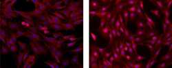
- Immunocytochemistry of fixed and permeabilized rat oligodendroglioma cells using 5 µg/mL of Anti-ERK1/2 eFluor® 570 (red). Cells were untreated (left) or treated with 50 ng/mL of PMA for 15 minutes (right). Nuclei are stained with Dapi (blue), colocalization appears pink.
Supportive validation
- Submitted by
- Invitrogen Antibodies (provider)
- Main image
- Experimental details
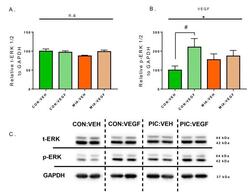
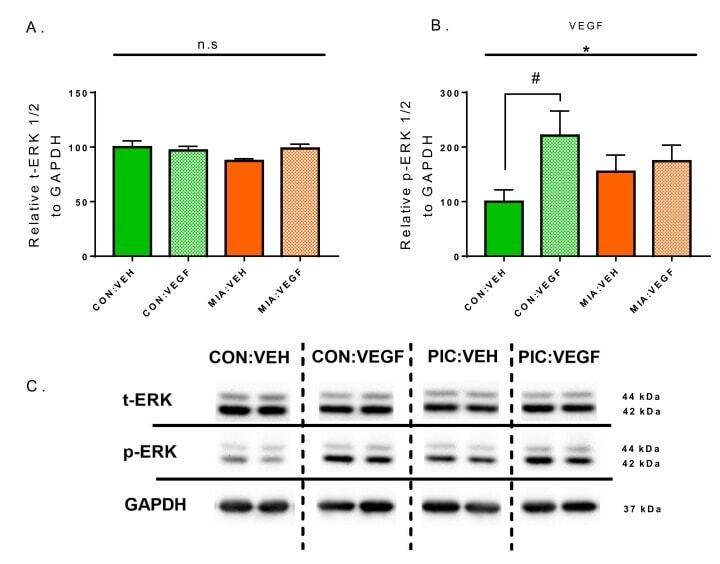
- Figure 3 Quantification of total ERK (t-ERK) and phospho-ERK (p-ERK) levels by Western blot. ( A ) Relative total ERK to GAPDH. ( B ) Relative p-ERK to GAPDH. ( C ) Representative Western blot images. Data are normalized to the CON:VEH group for comparison and presented as mean +- SEM. Statistical significances resulting from 2WAY-ANOVA are marked with * ( p < 0.05). Statistical significances resulting from Fisher''s LSD test are marked with # ( p < 0.05); n = 9-10/group.
- Submitted by
- Invitrogen Antibodies (provider)
- Main image
- Experimental details
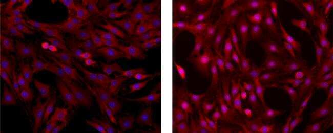
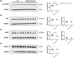
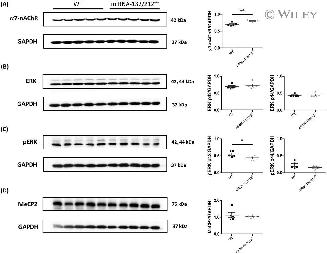
- 5 FIGURE The miRNA-132/212 deletion alters protein levels of alpha7-nAChR and pERK in mouse hippocampus. Total cell lysates from male mouse bilateral hippocampi were analysed by Western blot to determine the protein expression levels of alpha7-nAChR, ERK and pERK. A, Representative immunoblots and relative levels of alpha7-nAChR protein together with corresponding GAPDH levels, indicating significant increase in protein levels of alpha7-nAChR in the hippocampus of miRNA-132/212 -/- mice, compared with wild-type mice. B, Representative immunoblots and relative levels of ERK1 (p44) and ERK2 (p41) protein together with corresponding GAPDH levels in the hippocampus of miRNA-132/212 -/- mice. Analysis showed no changes in protein levels of ERK compared with wild-type mice. C, Representative immunoblots and relative levels of pERK and GAPDH levels indicating significant decrease in protein levels of pERK in the hippocampus of miRNA-132/212 -/- mice compared with wild-type mice. No significant differences were observed in protein levels of pERK. D, Representative immunoblots and relative levels of MeCP2 protein together with corresponding GAPDH levels, indicating no significant differences in protein levels of MeCP2 in the hippocampus of miRNA-132/212 -/- mice, compared with wild-type mice. Results are shown relative to GAPDH. Data are expressed as mean +- SD. n = 5 per group. WT, wild type; kDa, kilodalton. Please see details for the statistics on the main text
- Submitted by
- Invitrogen Antibodies (provider)
- Main image
- Experimental details
- FIGURE 5 The miRNA-132/212 deletion alters protein levels of alpha7-nAChR and pERK in mouse hippocampus. Total cell lysates from male mouse bilateral hippocampi were analysed by Western blot to determine the protein expression levels of alpha7-nAChR, ERK and pERK. A, Representative immunoblots and relative levels of alpha7-nAChR protein together with corresponding GAPDH levels, indicating significant increase in protein levels of alpha7-nAChR in the hippocampus of miRNA-132/212 -/- mice, compared with wild-type mice. B, Representative immunoblots and relative levels of ERK1 (p44) and ERK2 (p41) protein together with corresponding GAPDH levels in the hippocampus of miRNA-132/212 -/- mice. Analysis showed no changes in protein levels of ERK compared with wild-type mice. C, Representative immunoblots and relative levels of pERK and GAPDH levels indicating significant decrease in protein levels of pERK in the hippocampus of miRNA-132/212 -/- mice compared with wild-type mice. No significant differences were observed in protein levels of pERK. D, Representative immunoblots and relative levels of MeCP2 protein together with corresponding GAPDH levels, indicating no significant differences in protein levels of MeCP2 in the hippocampus of miRNA-132/212 -/- mice, compared with wild-type mice. Results are shown relative to GAPDH. Data are expressed as mean +- SD. n = 5 per group. WT, wild type; kDa, kilodalton. Please see details for the statistics on the main text

- Submitted by
- Invitrogen Antibodies (provider)
- Main image
- Experimental details
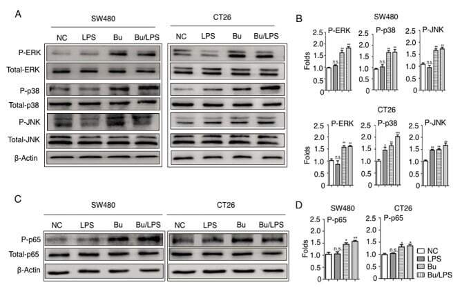
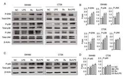
- Figure 5. Butyrate modifies the phosphorylation of ERK, p38, JNK and NF-kappaB p65 in colon cancer cells. The phosphorylation of ERK, p38, JNK and NF-kappaB p65 proteins in SW480 and CT26 cells treated with butyrate and/or LPS were detected using a western blot analysis assay. (A) Representative western blot analysis images of P-ERK/ERK, P-p38/p38 and P-JNK/JNK are presented. (B) The ratios of P-ERK/ERK, P-p38/p38 and P-JNK/JNK protein intensities were calculated. (C) Representative western blot analysis images of P-NF-kappaB p65/NF-kappaB p65 protein expression in cells are presented. (D) P-NF-kappaB p65/p65 protein intensities were calculated. beta-actin was used as the control. Experiments were performed in triplicate and data are expressed as the mean +- standard deviation. Compared with the NC group, *P