Explore
 Validate
 Learn
Learn Western blot
 Immunohistochemistry
Antibody data
- Antibody Data
- Antigen structure
- References [0]
- Comments [0]
- Validations
- Immunohistochemistry [3]
- Flow cytometry [1]
Submit
Validation data
Reference
Comment
Report error
- Product number
- NBP2-67359 - Provider product page

- Provider
- Novus Biologicals
- Product name
- Rabbit Monoclonal ERK1/2 Antibody
- Antibody type
- Monoclonal
- Description
- Protein A purified.
- Reactivity
- Human, Mouse
- Host
- Rabbit
- Isotype
- IgG
- Vial size
- 100 ul
- Storage
- Store at 4C short term. Aliquot and store at -20C long term. Avoid freeze-thaw cycles.
No comments: Submit comment
Supportive validation
- Submitted by
- Novus Biologicals (provider)
- Main image

- Experimental details
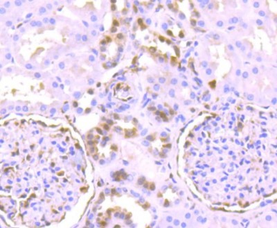
- Immunohistochemistry-Paraffin: ERK1/2 [p Thr202, p Thr185] Antibody (SZ2-4) [NBP2-67359] - Analysis of paraffin-embedded human kidney tissue using anti-Phospho-Erk1(T202)+Erk2(T185) antibody. Counter stained with hematoxylin.
- Submitted by
- Novus Biologicals (provider)
- Main image
- Experimental details
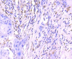
- Immunohistochemistry-Paraffin: ERK1/2 [p Thr202, p Thr185] Antibody (SZ2-4) [NBP2-67359] - Analysis of paraffin-embedded human lung cancer tissue using anti-Phospho-Erk1(T202)+Erk2(T185) antibody. Counter stained with hematoxylin.
- Submitted by
- Novus Biologicals (provider)
- Main image
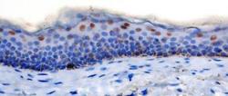
- Experimental details
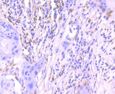
- Immunohistochemistry-Paraffin: ERK1/2 [p Thr202, p Thr185] Antibody (SZ2-4) [NBP2-67359] - Staining in canine epidermis. Antigen retrieval: citrate buffer (pH 6.0) for 10 minutes at 121C. Blocking of nonspecific reactions in 3% hydrogen peroxide in methanol at room temperature for 5min, followed by 8% skimmed milk at 37C for 40min. Primary antibody dilution: 1:100 and Secondary antibody: Dako EnVision + System. Reaction products were visualized with 0.05% 3.3'-diaminobenzidine (DAB) and 0.03% hydrogen peroxide in Tris-hydrochloric buffer and then counterstained with Mayer's hematoxylin. IHC-P image submitted by verified customer review.

Supportive validation
- Submitted by
- Novus Biologicals (provider)
- Main image
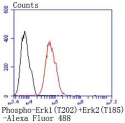
- Experimental details
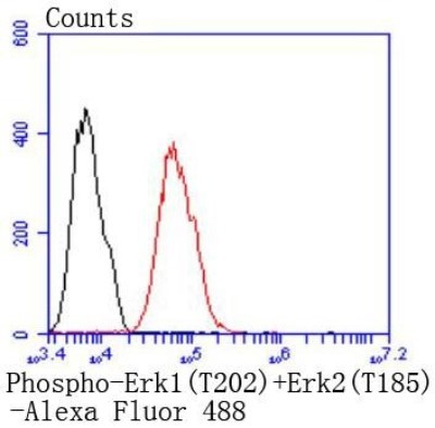
- Flow Cytometry: ERK1/2 [p Thr202, p Thr185] Antibody (SZ2-4) [NBP2-67359] - Analysis of MCF-7 cells with Phospho-Erk1(T202)+Erk2(T185) antibody at 1:50 dilution (red) compared with an unlabelled control (cells without incubation with primary antibody; black). Alexa Fluor 488-conjugated goat anti rabbit IgG was used as secondary antibody.