Explore
 Validate
 Learn
Learn Western blot
Antibody data
- Antibody Data
- Antigen structure
- References [0]
- Comments [0]
- Validations
- Western blot [3]
- Immunocytochemistry [1]
- Other assay [1]
Submit
Validation data
Reference
Comment
Report error
- Product number
- PA5-13036 - Provider product page

- Provider
- Invitrogen Antibodies
- Product name
- Phospho-ERK1 (Thr202, Tyr205) Polyclonal Antibody
- Antibody type
- Polyclonal
- Antigen
- Synthetic peptide
- Description
- This antibody is predicted to react with canine, mouse and rat based on sequence homology.
- Reactivity
- Human
- Host
- Rabbit
- Isotype
- IgG
- Vial size
- 400 μL
- Concentration
- 0.5 mg/mL
- Storage
- Store at 4°C short term. For long term storage, store at -20°C, avoiding freeze/thaw cycles.
No comments: Submit comment
Supportive validation
- Submitted by
- Invitrogen Antibodies (provider)
- Main image

- Experimental details
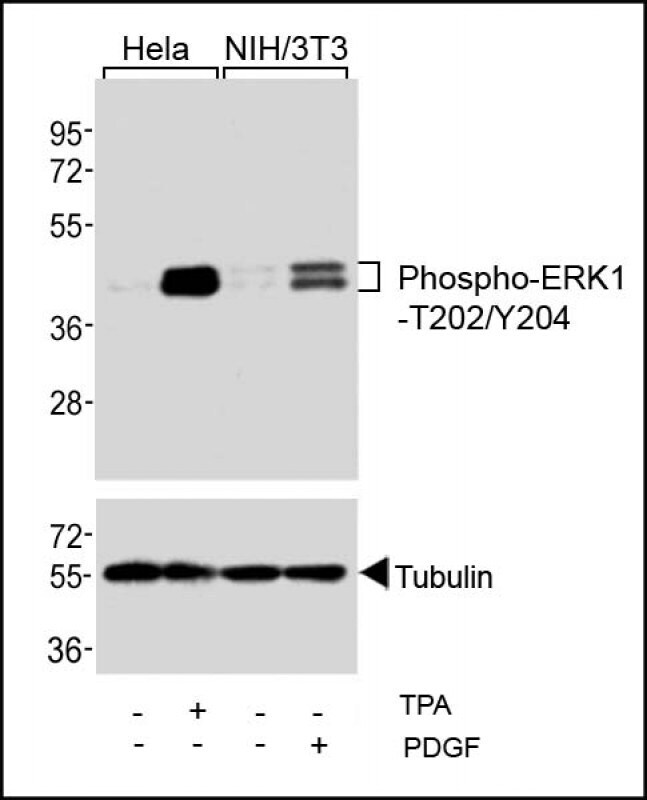
- Western blot analysis of Phospho-ERK1 (Thr202, Tyr205) in extracts from Hela and NIH/3T3 cells. Samples were incubated with Phospho-ERK1 (Thr202, Tyr205) polyclonal antibody (Product # PA5-13036). Phospho-ERK1-T202/Y204 Antibody (upper) or Tubulin (lower).
- Submitted by
- Invitrogen Antibodies (provider)
- Main image
- Experimental details
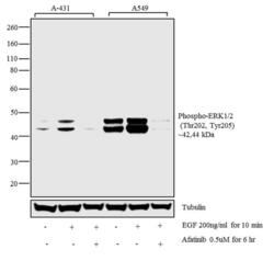
- Western blot analysis was performed on whole cell extracts (30 µg) of A-431 (1), A-431 treated with EGF (200 ng/mL for 10 minutes) (2), A-431 treated with Afatinib followed by EGF (0.5 uM of Afatinib for 6hrs, 200 ng/mL for 10 minutes) (3), A549 (4) and A549 treated with EGF (200 ng/mL for 10 minutes) (5). The blot was probed with Anti-Phospho-ERK1/ERK2 (Thr202, Tyr205) Rabbit Polyclonal Antibody (Product# PA5-13036, 1:1000 dilution) and detected by chemiluminescence using Goat anti-Rabbit IgG (Heavy Chain) Superclonal™ Secondary Antibody, HRP conjugate (Product # A27036, 0.25 µg/mL, 1:4000 dilution). 44, 42 kDa band corresponding to Phospho-ERK1/ERK2 (Thr202, Tyr205) was detected EGF treatment across cell lines tested and pre-treatment with Afatinib (antagonist) resulted in inhibition of Phospho-ERK1/ERK2 (Thr202, Tyr205) in A-431 cell line upon EGF treatment. Known quantity of protein samples were electrophoresed using Novex® NuPAGE® 4-12 % Bis-Tris gel (Product # NP0321BOX), XCell SureLock™ Electrophoresis System (Product # EI0002) and Novex Protein Standard (Product # LC5800). Resolved proteins were then transferred onto a nitrocellulose membrane by wet transfer method. The membrane was probed with the relevant primary and secondary Antibody following blocking with 5 % skimmed milk. Chemiluminescent detection was performed using Pierce™ ECL Western Blotting Substrate (Product # 32106).

- Submitted by
- Invitrogen Antibodies (provider)
- Main image
- Experimental details
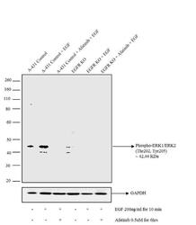
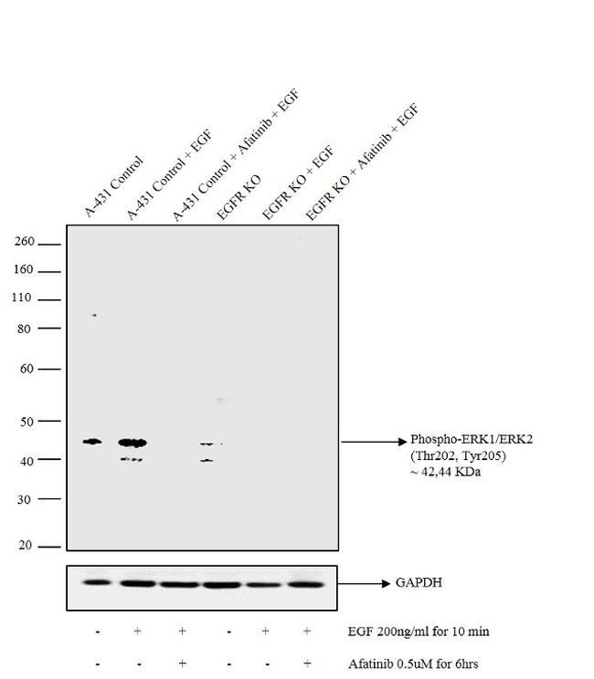
- Western blot analysis was performed by loading 30 µg of A-431 (Lane 1), A-431 treated with EGF (200 ng/mL for 10 minutes) (Lane 2), A-431 treated with Afatinib followed by EGF (0.5 uM of Afatinib for 6hrs, 200 ng/mL for 10 minutes) (Lane 3), A-431 EGFR KO (Lane 4) A-431 EGFR KO treated with EGF (200 ng/mL for 10 minutes) (Lane 5), A-431 EGFR KO treated with Afatinib followed by EGF (0.5 uM of Afatinib for 6hrs, 200 ng/mL for 10 minutes) (Lane 6) whole cell extracts using Novex® NuPAGE® 4-12 % Bis-Tris gel (Product # NP0321BOX), XCell SureLock™ Electrophoresis System (Product # EI0002) and Novex Protein Standard (Product # LC5800). Proteins were transferred to a nitrocellulose membrane using iBlot® Dry Blotting System (IB21001) and blocked with 5% skim milk for 1 hour at room temperature. The blot was probed with Anti-Phospho-ERK1/ERK2 (Thr202, Tyr205) Rabbit Polyclonal Antibody (Product# PA5-13036, 1:1000 dilution) and detected by chemiluminescence using Goat anti-Rabbit IgG (Heavy Chain) Superclonal™ Secondary Antibody, HRP conjugate (Product # A27036, 0.25 µg/mL, 1:4000 dilution). 44, 42 kDa band corresponding to Phospho-ERK1/ERK2 (Thr202, Tyr205) was detected in EGF treatment in control cell line (lane1 and 2) and not in EGFR knockout cell line (lane 4 and 5). Pre-treatment with Afatinib (antagonist) resulted in inhibition of Phospho-ERK1/ERK2 (Thr202, Tyr205) upon EGF treatment (lane 3 and 6).
Supportive validation
- Submitted by
- Invitrogen Antibodies (provider)
- Main image
- Experimental details
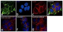
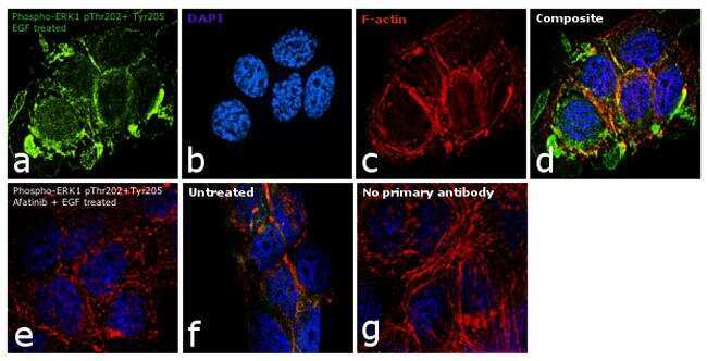
- Immunofluorescence analysis of Phospho-ERK1 (Thr202, Tyr205) was performed using 90% confluent log phase A-431 cells treated with 200 ng/mL of EGF for 10 minutes. The cells were fixed with 4% paraformaldehyde for 10 minutes, permeabilized with 0.1% Triton™ X-100 for 15 minutes, and blocked with 1% BSA for 1 hour at room temperature. The cells were labeled with Phospho-ERK1 (Thr202, Tyr205) Rabbit Polyclonal Antibody (Product # PA5-13036) at 5 µg in 0.1% BSA and incubated overnight at 4 degree Celsius and then labelled with Goat anti-Rabbit IgG (Heavy Chain) Superclonal™ Secondary Antibody, Alexa Fluor® 488 conjugate (Product # A27034) at a dilution of 1:2000 for 45 minutes at room temperature (Panel a: green). Nuclei (Panel b: blue) were stained with SlowFade® Gold Antifade Mountant with DAPI (Product # S36938). F-actin (Panel c: red) was stained with Rhodamine Phalloidin (Product # R415, 1:100). Panel d represents the merged image showing membrane and cytoplasmic localization. Panel e represents cells treated with antagonist, Afatinib (1µM for 6hrs) followed by EGF (200 ng/mL for 10 minutes), showing no signal. Panel f shows untreated cells with nuclear and cytoplasmic staining. Panel g represents control cells with no primary antibody to assess background. The images were captured at 60X magnification.
Supportive validation
- Submitted by
- Invitrogen Antibodies (provider)
- Main image

- Experimental details
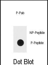
- Dot blot analysis of Phospho-ERK1 (Thr202, Tyr205). Samples were incubated with Phospho-ERK1 (Thr202, Tyr205) polyclonal antibody (Product # PA5-13036) using a dilution of 0.5 µg/mL. 50 ng of Bisphospho-peptide or Non Phosphorylated peptide per dot were adsorbed on nitrocellulose membrane.