Explore
 Validate
 Learn
Learn Western blot
Antibody data
- Antibody Data
- Antigen structure
- References [0]
- Comments [0]
- Validations
- Western blot [1]
- Immunohistochemistry [7]
Submit
Validation data
Reference
Comment
Report error
- Product number
- LS-C99745 - Provider product page

- Provider
- LSBio
- Proper citation
- LifeSpan Cat#LS-C99745, RRID:AB_2285966
- Product name
- SGK3 Antibody (aa1-30) LS-C99745
- Antibody type
- Polyclonal
- Description
- Ammonium sulfate precipitation
- Reactivity
- Human
- Host
- Rabbit
- Storage
- Maintain refrigerated at 2°C to 8°C for up to 6 months. For long term storage store at -20°C.
No comments: Submit comment
Enhanced validation
- Submitted by
- LSBio (provider)
- Enhanced method
- Genetic validation
- Main image
- Experimental details
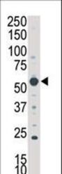
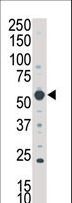
- Western blot of anti-SKG3 antibody in A375 cell lysate. SGK3 (arrow) was detected using purified antibody. Secondary HRP-anti-rabbit was used for signal visualization with chemiluminescence.
Enhanced validation
- Submitted by
- LSBio (provider)
- Enhanced method
- Genetic validation
- Main image

- Experimental details
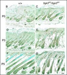
- IHC detection of SGK3 protein on the paraffin sections of the WT (left) and YPC (right) mice at P0 (B and C), P3 (D and E), and P5 (F and G) skin. Positive signals were observed in the cytoplasm of the hair follicle keratinocytes, especially in hair bulb, ORS, IRS, cuticle/cortex and bulge, or sebaceous glands. Some differences between the WT and YPC, for example, the expression in bulb keratinocytes were observed at P3 and P5. Scale bar, 50.
- Submitted by
- LSBio (provider)
- Enhanced method
- Genetic validation
- Main image
- Experimental details
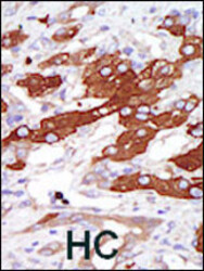
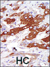
- Formalin-fixed and paraffin-embedded human cancer tissue reacted with the primary antibody, which was peroxidase-conjugated to the secondary antibody, followed by DAB staining. This data demonstrates the use of this antibody for immunohistochemistry; clinical relevance has not been evaluated. BC = breast carcinoma; HC = hepatocarcinoma.
- Submitted by
- LSBio (provider)
- Enhanced method
- Genetic validation
- Main image

- Experimental details
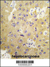
- Formalin-fixed and paraffin-embedded human hepatocarcinoma tissue reacted with SGK3 Antibody , which was peroxidase-conjugated to the secondary antibody, followed by DAB staining. This data demonstrates the use of this antibody for immunohistochemistry; clinical relevance has not been evaluated.

- Submitted by
- LSBio (provider)
- Enhanced method
- Genetic validation
- Main image

- Experimental details
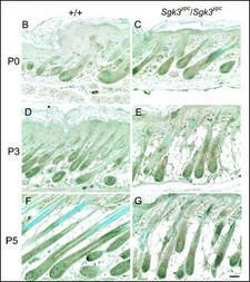
- IHC detection of SGK3 protein on the paraffin sections of the WT (left) and YPC (right) mice at P0 (B and C), P3 (D and E), and P5 (F and G) skin. Positive signals were observed in the cytoplasm of the hair follicle keratinocytes, especially in hair bulb, ORS, IRS, cuticle/cortex and bulge, or sebaceous glands. Some differences between the WT and YPC, for example, the expression in bulb keratinocytes were observed at P3 and P5. Scale bar, 50.

- Submitted by
- LSBio (provider)
- Main image

- Experimental details
- Formalin-fixed and paraffin-embedded human cancer tissue reacted with the primary antibody, which was peroxidase-conjugated to the secondary antibody, followed by DAB staining. This data demonstrates the use of this antibody for immunohistochemistry; clinical relevance has not been evaluated. BC = breast carcinoma; HC = hepatocarcinoma.

- Submitted by
- LSBio (provider)
- Main image

- Experimental details
- Formalin-fixed and paraffin-embedded human hepatocarcinoma tissue reacted with SGK3 Antibody , which was peroxidase-conjugated to the secondary antibody, followed by DAB staining. This data demonstrates the use of this antibody for immunohistochemistry; clinical relevance has not been evaluated.
- Submitted by
- LSBio (provider)
- Main image

- Experimental details
- IHC detection of SGK3 protein on the paraffin sections of the WT (left) and YPC (right) mice at P0 (B and C), P3 (D and E), and P5 (F and G) skin. Positive signals were observed in the cytoplasm of the hair follicle keratinocytes, especially in hair bulb, ORS, IRS, cuticle/cortex and bulge, or sebaceous glands. Some differences between the WT and YPC, for example, the expression in bulb keratinocytes were observed at P3 and P5. Scale bar, 50.