Explore
 Validate
 Learn
Learn Western blot
Antibody data
- Antibody Data
- Antigen structure
- References [0]
- Comments [0]
- Validations
- Western blot [4]
- Immunocytochemistry [4]
- Immunohistochemistry [1]
- Flow cytometry [1]
Submit
Validation data
Reference
Comment
Report error
- Product number
- MA5-49360 - Provider product page

- Provider
- Invitrogen Antibodies
- Product name
- GPNMB Recombinant Rabbit Monoclonal Antibody (PSH0-82)
- Antibody type
- Monoclonal
- Antigen
- Recombinant full-length protein
- Description
- Sequence Similarities: 98% Mouse/Rat. Tissue Specificity: Widely expressed, but very low expression, if any, in the brain (PubMed:12609765, PubMed:16609006). Expressed in the epidermis with higher levels in melanocytes compared with keratinocytes and Langerhans cells (at protein level) (PubMed:29336782). Expressed in peripheral blood, but not bone marrow mononuclear cells (PubMed:12609765). Expressed in tissue macrophages, including liver Kuppfer cells and lung alveolar macrophages, in podocytes and in some cells of the ciliary body of the eye (at protein level) (PubMed:16489096). May be overexpressed in various cancers, including melanoma and glioblastoma multiforme (PubMed:7814155, PubMed:16489096, PubMed:16609006). Positive Control: L929 cell lysate, A375 cell lysate, U-937 cell lysate, rat heart tissue lysate, B16F1 cell lysates, human lung tissue, L929. Subcellular Location: Cell membrane, Melanosome membrane, Early endosome membrane. Predicted band size: 64 kDa.
- Reactivity
- Human, Mouse, Rat
- Host
- Rabbit
- Isotype
- IgG
- Antibody clone number
- PSH0-82
- Vial size
- 100 μL
- Concentration
- 1 mg/mL
- Storage
- Store at 4°C short term. For long term storage, store at -20°C, avoiding freeze/thaw cycles.
No comments: Submit comment
Supportive validation
- Submitted by
- Invitrogen Antibodies (provider)
- Main image
- Experimental details
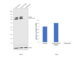
- Knockdown of GPNMB was achieved by transfecting SK-MEL-5 with GPNMB specific siRNAs (Silencer® select Product # s20461, s20462). Western blot analysis (Fig. a) was performed using whole cell extracts from the GPNMB knockdown cells (lane 3), non-targeting scrambled siRNA transfected cells (lane 2) and untransfected cells (lane 1). The blot was probed with GPNMB Rabbit Recombinant Monoclonal Antibody (PSH0-82) (Product # MA5-49360, 1:2,000 dilution ) and Goat anti-Rabbit IgG (Heavy Chain) Superclonal™ Recombinant Secondary Antibody, HRP (Product # A27036, 1:20,000 dilution). Densitometric analysis of this western blot is shown in histogram (Fig. b). Loss of signal upon siRNA mediated knock down confirms that antibody is specific to GPNMB.

- Submitted by
- Invitrogen Antibodies (provider)
- Main image
- Experimental details
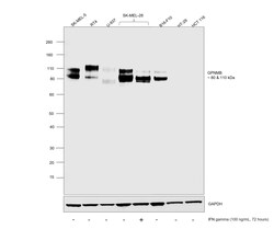
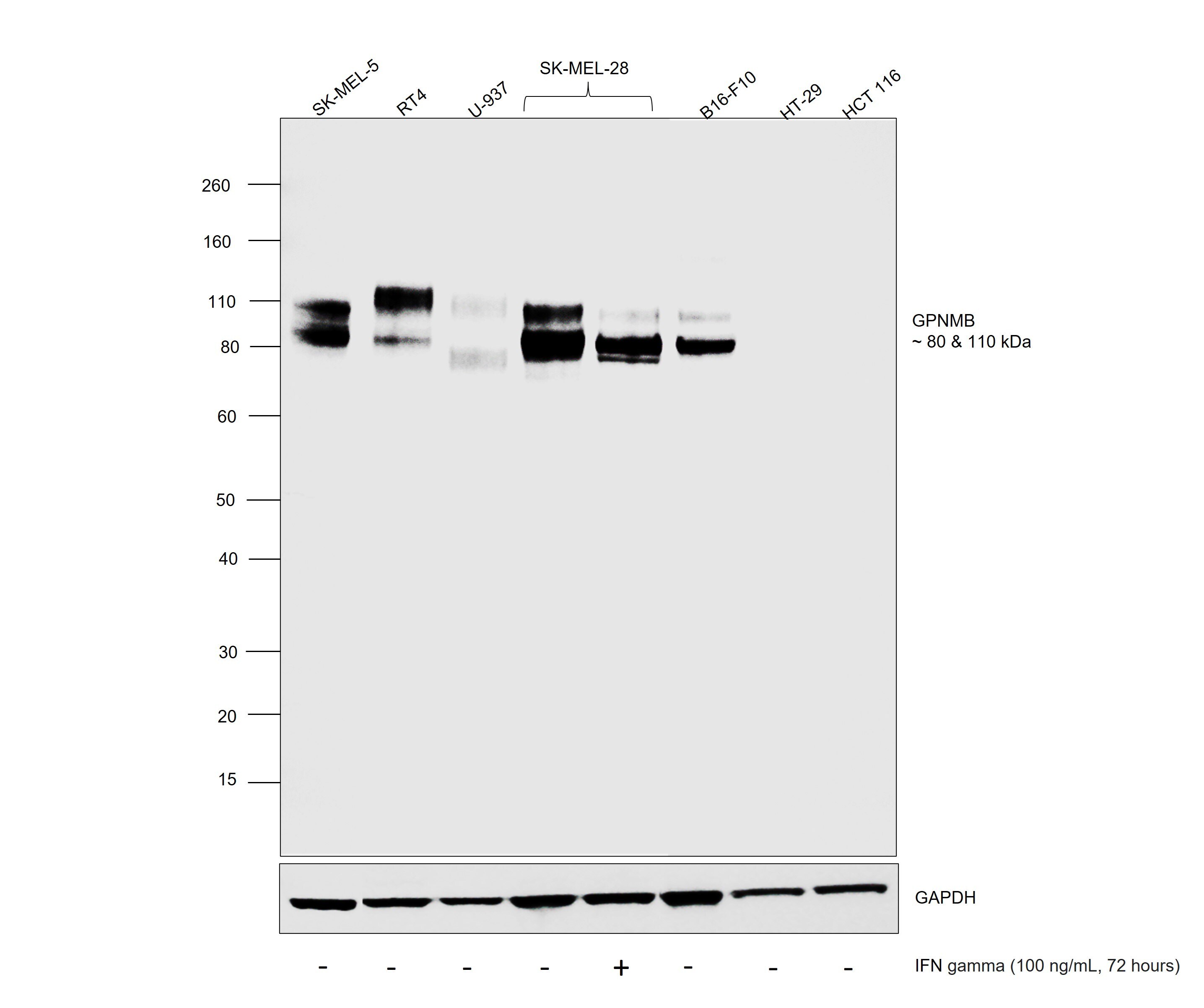
- Western blot was performed using GPNMB Rabbit Recombinant Monoclonal Antibody (PSH0-82) (Product # MA5-49360) and, 110 kDa and 80 kDa bands corresponding to GPNMB were observed across high expressing cell lines and not in low expressing cell lines tested. Whole cell extracts (30 µg lysate) of SK-MEL-5 (Lane 1), RT4 (Lane 2), U-937 (Lane 3), SK-MEL-28 untreated (Lane 4), SK-MEL-28 treated with IFN gamma at 100 ng/mL concentration for 72 hours (Lane 5), low expressing cell lines such as HT-29 (Lane 6) and HCT 116 (Lane 7) were electrophoresed using NuPAGE™ 4-12% Bis-Tris Protein Gel (Product # NP0321BOX), 10 well. Resolved proteins were then transferred onto a nitrocellulose membrane (Product # IB23001) by iBlot® 2 Dry Blotting System (Product # IB21001). The blot was probed with the primary antibody (1:2,000 dilution) and detected by chemiluminescence with Goat anti-Rabbit IgG (Heavy Chain) Superclonal™ Recombinant Secondary Antibody, HRP (Product # A27036, 1:20,000 dilution) using the iBright™ FL1500 Imaging System (Product # A44115). Chemiluminescent detection was performed using SuperSignal™ West Pico PLUS Chemiluminescent Substrate (Product # 34579). In lane 5, upon treatment with IFN gamma, GPNMB expression decreases in malignant melanoma as reported.
- Submitted by
- Invitrogen Antibodies (provider)
- Main image

- Experimental details
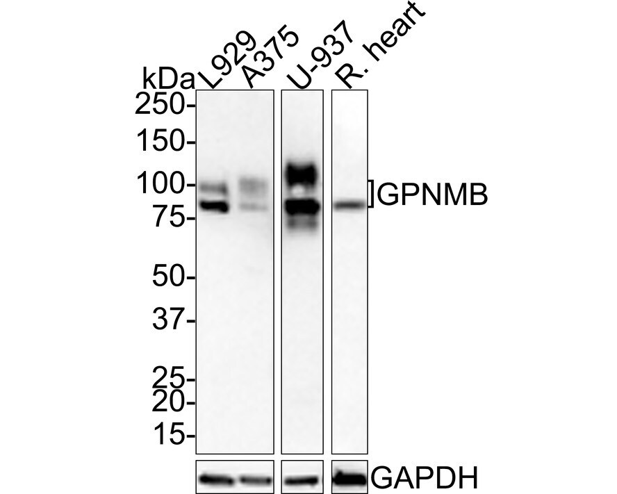
- Western blot was performed using GPNMB Rabbit Recombinant Monoclonal Antibody (PSH0-82) (Product # MA5-49360) and 80-120 kDa bands corresponding to GPNMB was observed across cell lines tested. Whole cell extracts (20 µg lysate) of L929 (Lane 1), A375 (Lane 2), U-937 (Lane 3) and Whole cell extracts (40 µg lysate) of Rat heart (Lane 4) were electrophoresed using 4-20% SDS-PAGE gel. Resolved proteins were transferred onto a PVDF membrane. The blot was blocked with 5% NFDM/TBST for 1 hour at room temperature, then probed with the primary antibody (1:1,000 dilution) for 2 hours at room temperature and detected by chemiluminescence with HRP labeled Goat anti-Rabbit IgG secondary antibody.
- Submitted by
- Invitrogen Antibodies (provider)
- Main image
- Experimental details
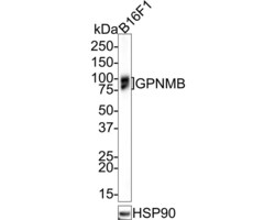
- Western blot was performed using GPNMB Rabbit Recombinant Monoclonal Antibody (PSH0-82) (Product # MA5-49360) and 80-120 kDa bands corresponding to GPNMB was observed across cell lines tested. Whole cell extract (20 µg lysate) of B16F1 was electrophoresed using 4-20% SDS-PAGE gel. Resolved proteins were transferred onto a PVDF membrane. The blot was blocked with 5% NFDM/TBST for 1 hour at room temperature, then probed with the primary antibody (1:1,000 dilution) for 2 hours at room temperature and detected by chemiluminescence with HRP labeled Goat anti-Rabbit IgG secondary antibody.

Supportive validation
- Submitted by
- Invitrogen Antibodies (provider)
- Main image
- Experimental details
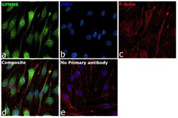
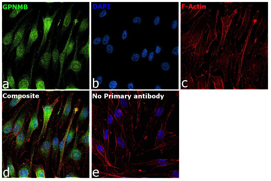
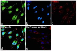
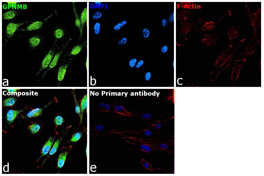
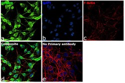
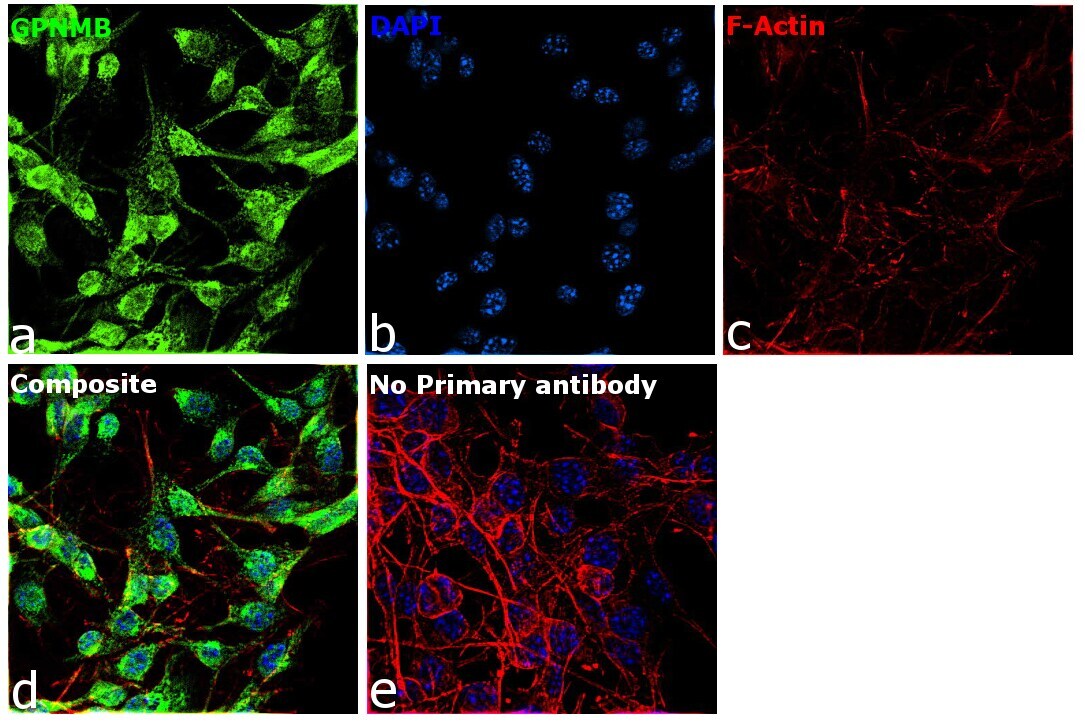
- Immunofluorescence analysis of GPNMB was performed using 70% confluent log phase SK-MEL-28 cells. The cells were fixed with 4% paraformaldehyde for 10 minutes, permeabilized with 0.1% Triton™ X-100 for 15 minutes, and blocked with 2% BSA for 1 hour at room temperature. The cells were labeled with GPNMB Rabbit Recombinant Monoclonal Antibody (PSH0-82) (Product # MA5-49360) at 1:100 dilution in 0.1% BSA, incubated at 4°C overnight and then labeled with Donkey anti-Rabbit IgG (H+L) Highly Cross-Adsorbed Secondary Antibody, Alexa Fluor™ Plus 488 (Product # A32790), (1:2,000 dilution), for 45 minutes at room temperature (Panel a: Green). Nuclei (Panel b: Blue) were stained with ProLong™ Diamond Antifade Mountant with DAPI (Product # P36962). F-actin (Panel c: Red) was stained with Rhodamine Phalloidin (Product # R415, 1:300 dilution). Panel d represents the merged image showing cell membrane and cytosolic localization. Panel e represents control cells with no primary antibody to assess background. The images were captured at 60X magnification.
- Submitted by
- Invitrogen Antibodies (provider)
- Main image
- Experimental details
- Immunofluorescence analysis of GPNMB using L929 cells. The cells were fixed 4% paraformaldehyde for 10 minutes, permeabilized with 0.05% Triton™ X-100 in PBS for 20 minutes, and blocked with 2% negative goat serum for 30 minutes at room temperature. The cells were labeled with GPNMB Rabbit Recombinant Monoclonal Antibody (PSH0-82) (Product # MA5-49360) at 1:100 dilution in 2% negative goat serum overnight at 4°C and then with iFluor™ 488 Goat Anti-Rabbit IgG H&L secondary antibody (1:1,000 dilution) for 1 hour at room temperature (green). Nuclear were stained with DAPI (blue). Beta tubulin (red) was stained at 1:200 dilution overnight at +4°C. iFluor™ 594 Goat Anti-Mouse IgG H&L was used as the secondary antibody at 1:1,000 dilution. The images were captured at 200X magnification.
- Submitted by
- Invitrogen Antibodies (provider)
- Main image
- Experimental details
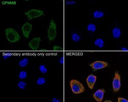
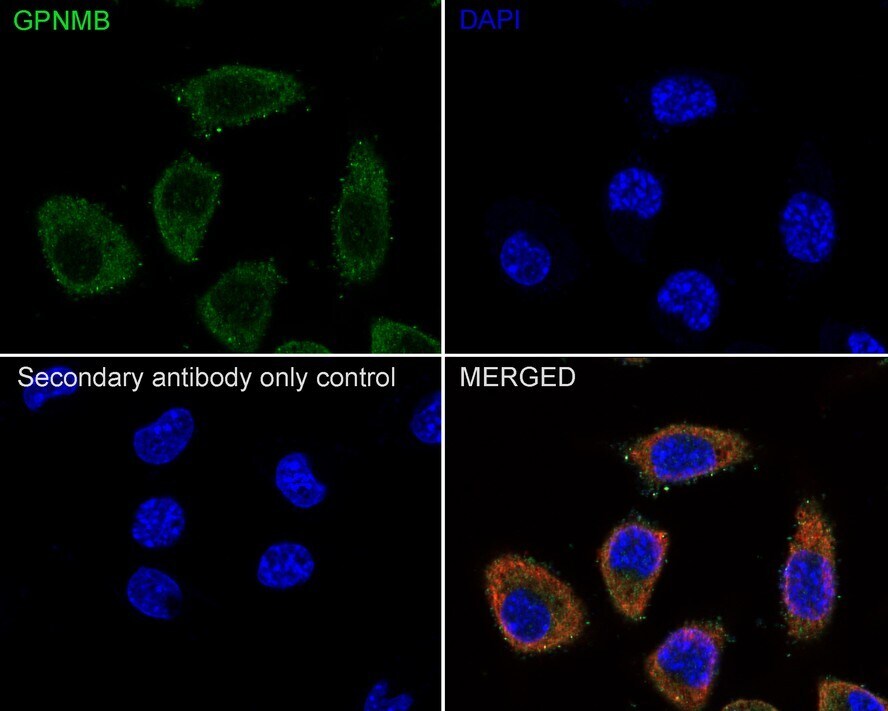
- Immunofluorescence analysis of GPNMB was performed using 70% confluent log phase SK-MEL-5 cells. The cells were fixed with 4% paraformaldehyde for 10 minutes, permeabilized with 0.1% Triton™ X-100 for 15 minutes, and blocked with 2% BSA for 1 hour at room temperature. The cells were labeled with GPNMB Rabbit Recombinant Monoclonal Antibody (PSH0-82) (Product # MA5-49360) at 1:100 dilution in 0.1% BSA, incubated at 4°C overnight and then labeled with Donkey anti-Rabbit IgG (H+L) Highly Cross-Adsorbed Secondary Antibody, Alexa Fluor™ Plus 488 (Product # A32790), (1:2,000 dilution), for 45 minutes at room temperature (Panel a: Green). Nuclei (Panel b: Blue) were stained with ProLong™ Diamond Antifade Mountant with DAPI (Product # P36962). F-actin (Panel c: Red) was stained with Rhodamine Phalloidin (Product # R415, 1:300 dilution). Panel d represents the merged image showing cell membrane and cytosolic localization. Panel e represents control cells with no primary antibody to assess background. The images were captured at 60X magnification.
- Submitted by
- Invitrogen Antibodies (provider)
- Main image
- Experimental details
- Immunofluorescence analysis of GPNMB was performed using 70% confluent log phase B16-F10 cells. The cells were fixed with 4% paraformaldehyde for 10 minutes, permeabilized with 0.1% Triton™ X-100 for 15 minutes, and blocked with 2% BSA for 1 hour at room temperature. The cells were labeled with GPNMB Rabbit Recombinant Monoclonal Antibody (PSH0-82) (Product # MA5-49360) at 1:100 dilution in 0.1% BSA, incubated at 4°C overnight and then labeled with Donkey anti-Rabbit IgG (H+L) Highly Cross-Adsorbed Secondary Antibody, Alexa Fluor™ Plus 488 (Product # A32790), (1:2,000 dilution), for 45 minutes at room temperature (Panel a: Green). Nuclei (Panel b: Blue) were stained with ProLong™ Diamond Antifade Mountant with DAPI (Product # P36962). F-actin (Panel c: Red) was stained with Rhodamine Phalloidin (Product # R415, 1:300 dilution). Panel d represents the merged image showing cell membrane and cytosolic localization. Panel e represents control cells with no primary antibody to assess background. The images were captured at 60X magnification.
Supportive validation
- Submitted by
- Invitrogen Antibodies (provider)
- Main image
- Experimental details
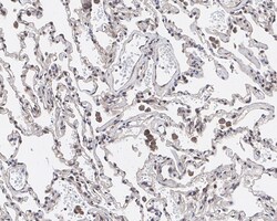
- Immunohistochemical analysis of GPNMB on formalin-fixed paraffin-embedded human lung tissue. The section was pre-treated using heat mediated antigen retrieval with Tris-EDTA buffer (pH 9.0) for 20 minutes. The tissue was blocked in 1% BSA for 20 minutes at room temperature, then probed with GPNMB Rabbit Recombinant Monoclonal Antibody (PSH0-82) (Product # MA5-49360) at 1:200 dilution for 1 hour at room temperature. HRP conjugated compact polymer system and DAB chromogen were used as the detection system, followed by counterstaining with hematoxylin. The slide was mounted with DPX and the image was captured at 200X magnification.

Supportive validation
- Submitted by
- Invitrogen Antibodies (provider)
- Main image
- Experimental details
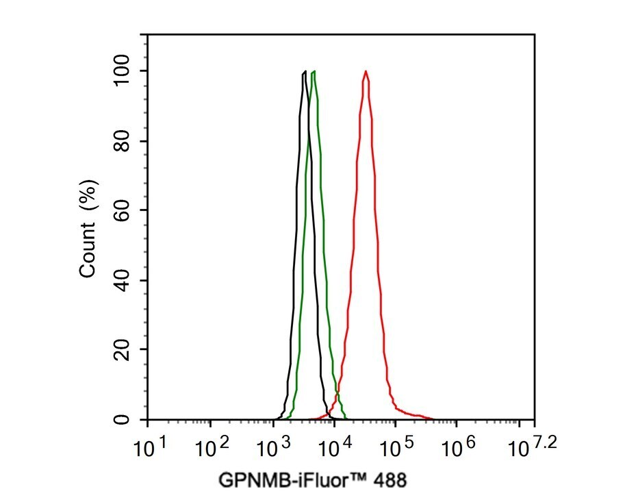
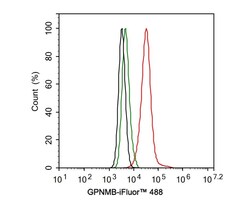
- Flow Cytometry analysis of GPNMB in L929 cells. The cells were fixed and permeabilized and then stained with GPNMB Rabbit Recombinant Monoclonal Antibody (PSH0-82) (Product # MA5-49360) at 1 µg/mL (red) and Rabbit IgG Isotype Control (green). After incubation of the primary antibody at 4°C for an hour, the cells were stained with a iFluor™ 488 Goat anti-Rabbit IgG Secondary antibody at 1:1,000 dilution for 30 minutes at 4°C. Unlabelled sample was used as a control (cells without incubation with primary antibody; black).