 Explore
 Validate
 Learn
LearnMA5-15858
antibody from Invitrogen Antibodies
Targeting: SMN1
BCD541, GEMIN1, SMA, SMA@, SMA1, SMA2, SMA3, SMNT, TDRD16A
 Western blot
 ELISA
ELISA Immunocytochemistry
Antibody data
- Antibody Data
- Antigen structure
- References [1]
- Comments [0]
- Validations
- Immunocytochemistry [4]
- Immunohistochemistry [2]
Submit
Validation data
Reference
Comment
Report error
- Product number
- MA5-15858 - Provider product page

- Provider
- Invitrogen Antibodies
- Product name
- SMN1 Monoclonal Antibody (5H1)
- Antibody type
- Monoclonal
- Antigen
- Purifed from natural sources
- Description
- MA5-15858 targets SMN1 in indirect ELISA, IF, IHC, and WB applications and shows reactivity with Human and Non-human primate samples. The MA5-15858 immunogen is purified recombinant fragment of human SMN1 expressed in E. Coli. . MA5-15858 detects SMN1 which has a predicted molecular weight of approximately 39kDa.
- Reactivity
- Human
- Host
- Mouse
- Isotype
- IgG
- Antibody clone number
- 5H1
- Vial size
- 100 μL
- Concentration
- Conc. not determined
- Storage
- Store at 4°C short term. For long term storage, store at -20°C, avoiding freeze/thaw cycles.
Submitted references Tumor Suppressor Analysis in CML.
Herrmann O, Schemionek M
Methods in molecular biology (Clifton, N.J.) 2016;1465:87-94
Methods in molecular biology (Clifton, N.J.) 2016;1465:87-94
No comments: Submit comment
Supportive validation
- Submitted by
- Invitrogen Antibodies (provider)
- Main image
- Experimental details
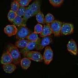
- Immunofluorescence analysis of HepG2 cells using SMN1 monoclonal antibody (Product # MA5-15858) (Green). Blue: DRAQ5 fluorescent DNA dye. Red: actin filaments have been labeled with phalloidin.

- Submitted by
- Invitrogen Antibodies (provider)
- Main image
- Experimental details
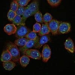
- Immunofluorescence analysis of HepG2 cells using SMN1 monoclonal antibody (Product # MA5-15858) (Green). Blue: DRAQ5 fluorescent DNA dye. Red: actin filaments have been labeled with phalloidin.
- Submitted by
- Invitrogen Antibodies (provider)
- Main image

- Experimental details
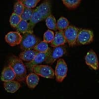
- Immunofluorescence analysis of HepG2 cells using SMN1 monoclonal antibody (Product # MA5-15858) (Green). Blue: DRAQ5 fluorescent DNA dye. Red: actin filaments have been labeled with phalloidin.

- Submitted by
- Invitrogen Antibodies (provider)
- Main image

- Experimental details
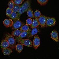
- Immunofluorescence analysis of HepG2 cells using SMN1 monoclonal antibody (Product # MA5-15858) (Green). Blue: DRAQ5 fluorescent DNA dye. Red: actin filaments have been labeled with phalloidin.
Supportive validation
- Submitted by
- Invitrogen Antibodies (provider)
- Main image
- Experimental details
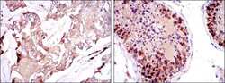
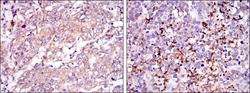
- Immunohistochemical analysis of paraffin-embedded breast cancer tissues (left) and testis tissues (right) using SMN1 monoclonal antibody (Product # MA5-15858) followed with DAB staining.
- Submitted by
- Invitrogen Antibodies (provider)
- Main image
- Experimental details
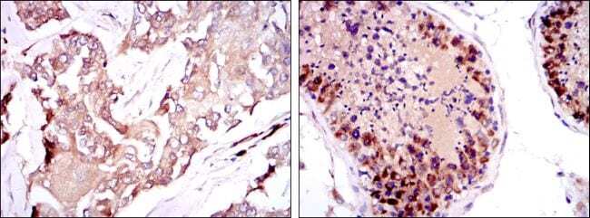
- Immunohistochemical analysis of paraffin-embedded stomach cancer tissues (left) and brain tumor (right) using SMN1 monoclonal antibody (Product # MA5-15858) followed with DAB staining.
