Explore
 Validate
 Learn
Learn Western blot
Antibody data
- Antibody Data
- Antigen structure
- References [1]
- Comments [0]
- Validations
- Western blot [3]
- Immunohistochemistry [1]
Submit
Validation data
Reference
Comment
Report error
- Product number
- AP1809a - Provider product page

- Provider
- Abcepta
- Proper citation
- Abgent Cat#AP1809a, RRID:AB_2274725
- Product name
- ATG4B Antibody (N-term)
- Antibody type
- Polyclonal
- Antigen
- Synthetic peptide
- Description
- Purified Rabbit Polyclonal Antibody (Pab)
- Reactivity
- Human, Mouse
- Host
- Rabbit
- Isotype
- IgG
- Vial size
- 400 µl
- Concentration
- 2 mg/ml
- Storage
- Maintain refrigerated at 2-8°C for up to 6 months. For long term storage store at -20°C in small aliquots to prevent freeze-thaw cycles.
Submitted references Nuclear expression of E2F4 induces cell death via multiple pathways in normal human intestinal epithelial crypt cells but not in colon cancer cells.
Garneau H, Alvarez L, Paquin MC, Lussier C, Rancourt C, Tremblay E, Beaulieu JF, Rivard N
American journal of physiology. Gastrointestinal and liver physiology 2007 Oct;293(4):G758-72
American journal of physiology. Gastrointestinal and liver physiology 2007 Oct;293(4):G758-72
No comments: Submit comment
Supportive validation
- Submitted by
- Abcepta (provider)
- Main image

- Experimental details
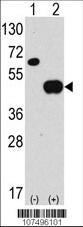
- Western blot analysis of anti-hAPG4B-R31 Pab (Cat. #AP1809a) in 293 cell line lysates transiently transfected with the ATG4B gene (2ug/lane). hAPG4B-R31(arrow) was detected using the purified Pab.
- Primary Ab dilution
- 1:1000
- Submitted by
- Abcepta (provider)
- Main image
- Experimental details
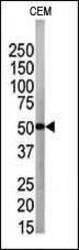
- The anti-APG4B Pab (Cat. #AP1809a) is used in Western blot to detect APG4B in CEM tissue lysate
- Primary Ab dilution
- 1:1000

- Submitted by
- Abcepta (provider)
- Main image
- Experimental details
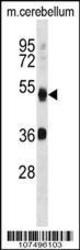
- APG4B Antibody (R31) (Cat. #AP1809a) western blot analysis in mouse cerebellum tissue lysates (35ug/lane).This demonstrates the APG4B antibody detected the APG4B protein (arrow).
- Primary Ab dilution
- 1:1000

Supportive validation
- Submitted by
- Abcepta (provider)
- Main image
- Experimental details
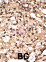
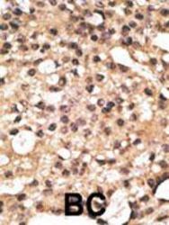
- "Formalin-fixed and paraffin-embedded human cancer tissue reacted with the primary antibody, which was peroxidase-conjugated to the secondary antibody, followed by DAB staining. This data demonstrates the use of this antibody for immunohistochemistry; clinical relevance has not been evaluated. BC = breast carcinoma; HC = hepatocarcinoma."
- Primary Ab dilution
- 1:50~100