Explore
 Validate
 Learn
Learn Western blot
 Immunocytochemistry
Antibody data
- Antibody Data
- Antigen structure
- References [0]
- Comments [0]
- Validations
- Immunocytochemistry [2]
- Immunohistochemistry [1]
- Flow cytometry [1]
Submit
Validation data
Reference
Comment
Report error
- Product number
- 701882 - Provider product page

- Provider
- Invitrogen Antibodies
- Product name
- ATG4B Recombinant Rabbit Monoclonal Antibody (1HC6LC10)
- Antibody type
- Monoclonal
- Antigen
- Other
- Description
- This antibody is predicted to react with Mouse, Bovine and Chicken. Recombinant rabbit monoclonal antibodies are produced using in vitro expression systems. The expression systems are developed by cloning in the specific antibody DNA sequences from immunoreactive rabbits. Then, individual clones are screened to select the best candidates for production. The advantages of using recombinant rabbit monoclonal antibodies include: better specificity and sensitivity, lot-to-lot consistency, animal origin-free formulations, and broader immunoreactivity to diverse targets due to larger rabbit immune repertoire.
- Reactivity
- Human, Mouse
- Host
- Rabbit
- Isotype
- IgG
- Antibody clone number
- 1HC6LC10
- Vial size
- 100 μg
- Concentration
- 0.5 mg/mL
- Storage
- Store at 4°C short term. For long term storage, store at -20°C, avoiding freeze/thaw cycles.
No comments: Submit comment
Supportive validation
- Submitted by
- Invitrogen Antibodies (provider)
- Main image
- Experimental details
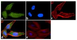
- Immunofluorescence was performed on fixed and permeabilized HeLa cells for detection of ATG4b using Anti-ATG4b Recombinant Rabbit Monoclonal Antibody (Product # 701882, 0.5-1 µg/mL) and labeled with Goat anti-Rabbit IgG (H+L) Superclonal Secondary Antibody, Alexa Fluor® 488 conjugate (Product # A27034, 0.4 µg/mL, 1:2500). Panel a) shows representative cells that were stained for detection and localization of ATG4b protein (green), Panel b) is stained for nuclei (blue) using SlowFade® Gold Antifade Mountant with DAPI (Product # S36938, 1:50). Panel c) represents cytoskeletal F-actin staining using Alexa Fluor® 594 Phalloidin (Product # A12381, 1:200). Panel d) is a composite image of Panels a, b and c clearly demonstrating cytoplasmic localization of ATG4b. Panel e) represents control cells with no primary Antibody to assess background.
- Submitted by
- Invitrogen Antibodies (provider)
- Main image

- Experimental details
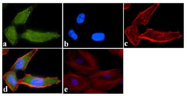
- Immunofluorescence was performed on fixed and permeabilized HeLa cells for detection of ATG4b using Anti-ATG4b Recombinant Rabbit Monoclonal Antibody (Product # 701882, 0.5-1 µg/mL) and labeled with Goat anti-Rabbit IgG (Heavy Chain) Superclonal Secondary Antibody, Alexa Fluor® 488 conjugate (Product # A27034, 0.4 µg/mL, 1:2500). Panel a) shows representative cells that were stained for detection and localization of ATG4b protein (green), Panel b) is stained for nuclei (blue) using SlowFade® Gold Antifade Mountant with DAPI (Product # S36938, 1:50). Panel c) represents cytoskeletal F-actin staining using Alexa Fluor® 594 Phalloidin (Product # A12381, 1:200). Panel d) is a composite image of Panels a, b and c clearly demonstrating cytoplasmic localization of ATG4b. Panel e) represents control cells with no primary Antibody to assess background.

Supportive validation
- Submitted by
- Invitrogen Antibodies (provider)
- Main image
- Experimental details
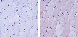
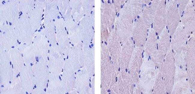
- Immunohistochemistry analysis of ATG4b showing staining in the cytoplasm of paraffin-embedded human skeletal muscle tissue (right) compared to a negative control without primary antibody (left). To expose target proteins, antigen retrieval was performed using 10mM sodium citrate (pH 6.0), microwaved for 8-15 min. Following antigen retrieval, tissues were blocked in 3% H2O2-methanol for 15 min at room temperature, washed with ddH2O and PBS, and then probed with a ATG4b Recombinant Rabbit Monoclonal Antibody (Product # 701882) diluted in 3% BSA-PBS at a dilution of 1:20 for 1 hour at 37ºC in a humidified chamber. Tissues were washed extensively in PBST and detection was performed using an HRP-conjugated secondary antibody followed by colorimetric detection using a DAB kit. Tissues were counterstained with hematoxylin and dehydrated with ethanol and xylene to prep for mounting.
Supportive validation
- Submitted by
- Invitrogen Antibodies (provider)
- Main image
- Experimental details
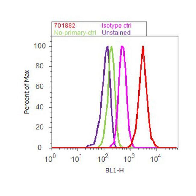
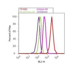
- Flow Cytometry analysis of ATG4b was performed on U-87 MG cells labeled with ABfinityª ATG4b Recombinant Rabbit Monoclonal Antibody (Product# 701882, 2 - 4ug/1M cells) or with Rabbit isotype control and detected with Goat anti-Rabbit IgG (H+L) Superclonalª Secondary Antibody, Alexa Fluor¨ 488 conjugate (Product # A27034, 0.4 ug/ml, 1:2500) as represented by the red and pink histograms respectively. The purple histogram represents unstained control cells and the green histogram represents no-primary-Antibody control. A representative 10,000 cells were acquired and analyzed for each sample using an Attune¨ Acoustic Focusing Cytometer (4468770).