 Explore
 Validate
 Learn
Learn Western blot
Antibody data
- Antibody Data
- Antigen structure
- References [0]
- Comments [0]
- Validations
- Western blot [5]
- Immunocytochemistry [1]
Submit
Validation data
Reference
Comment
Report error
- Product number
- 710915 - Provider product page

- Provider
- Invitrogen Antibodies
- Product name
- ATG4B Recombinant Polyclonal Antibody (1HCLC)
- Antibody type
- Polyclonal
- Antigen
- Other
- Description
- This antibody is predicted to react with monkey, mouse, rat, guinea pig and bovine based on sequence homology.
- Antibody clone number
- 1HCLC
- Concentration
- 0.5 mg/mL
No comments: Submit comment
Supportive validation
- Submitted by
- Invitrogen Antibodies (provider)
- Main image

- Experimental details
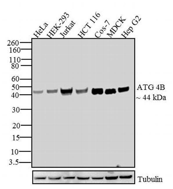
- Western blot analysis of ATG4B was performed by loading 30 µg of HeLa (lane 1), HEK 293 (lane 2), Jurkat (lane 3), HCT 116 (lane 4), COS 7 (lane 5), MDCK (lane 6) and HepG2 (lane 7) cell lysates using Novex® NuPAGE® 4-12 % Bis-Tris gel (Product # NP0321BOX), XCell SureLock™ Electrophoresis System (Product # EI0002), Novex® Sharp Pre-Stained Protein Standard (Product # LC5800) and iBlot® Dry Blotting System (Product # IB21001). Proteins were transferred to a nitrocellulose membrane and blocked with 5 % skim milk for 1 hour at room temperature on a rocking platform. ATG4B was detected at ~44 kDa using ATG 4B Recombinant Rabbit Polyclonal Antibody (Product # 710915) at 1-2 µg/mL in 5 % skim milk at 4°C overnight on a rocking platform. Goat anti-Rabbit IgG - HRP Secondary Antibody (Product # G-21234) at 1:5000 dilution was used and chemiluminescent detection was performed using Pierce™ ECL Western blotting Substrate (Product # 32106).
- Submitted by
- Invitrogen Antibodies (provider)
- Main image

- Experimental details
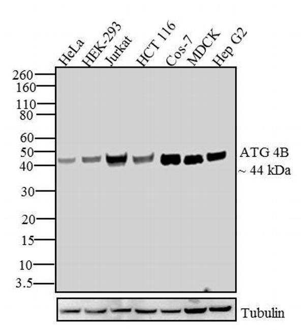
- Western blot analysis of ATG4B was performed by loading 30 µg of HeLa (lane 1), HEK 293 (lane 2), Jurkat (lane 3), HCT 116 (lane 4), COS 7 (lane 5), MDCK (lane 6) and HepG2 (lane 7) cell lysates using Novex® NuPAGE® 4-12 % Bis-Tris gel (Product # NP0321BOX), XCell SureLock™ Electrophoresis System (Product # EI0002), Novex® Sharp Pre-Stained Protein Standard (Product # LC5800) and iBlot® Dry Blotting System (Product # IB21001). Proteins were transferred to a nitrocellulose membrane and blocked with 5 % skim milk for 1 hour at room temperature on a rocking platform. ATG4B was detected at ~44 kDa using ATG 4B Recombinant Rabbit Polyclonal Antibody (Product # 710915) at 1-2 µg/mL in 5 % skim milk at 4°C overnight on a rocking platform. Goat anti-Rabbit IgG - HRP Secondary Antibody (Product # G-21234) at 1:5000 dilution was used and chemiluminescent detection was performed using Pierce™ ECL Western blotting Substrate (Product # 32106).
- Submitted by
- Invitrogen Antibodies (provider)
- Main image

- Experimental details
- Western blot analysis of ATG4B was performed by loading 30 µg of HeLa (lane 1), HEK 293 (lane 2), Jurkat (lane 3), HCT 116 (lane 4), COS 7 (lane 5), MDCK (lane 6) and HepG2 (lane 7) cell lysates using Novex® NuPAGE® 4-12 % Bis-Tris gel (Product # NP0321BOX), XCell SureLock™ Electrophoresis System (Product # EI0002), Novex® Sharp Pre-Stained Protein Standard (Product # LC5800) and iBlot® Dry Blotting System (Product # IB21001). Proteins were transferred to a nitrocellulose membrane and blocked with 5 % skim milk for 1 hour at room temperature on a rocking platform. ATG4B was detected at ~44 kDa using ATG 4B Recombinant Rabbit Polyclonal Antibody (Product # 710915) at 1-2 µg/mL in 5 % skim milk at 4°C overnight on a rocking platform. Goat anti-Rabbit IgG - HRP Secondary Antibody (Product # G-21234) at 1:5000 dilution was used and chemiluminescent detection was performed using Pierce™ ECL Western blotting Substrate (Product # 32106).

- Submitted by
- Invitrogen Antibodies (provider)
- Main image

- Experimental details
- Knockdown of ATG4B was achieved by transfecting MCF7 with ATG4B specific siRNAs (Silencer® select Product # s23244, s23246). Western blot analysis (Fig. a) was performed using whole cell extracts from the ATG4B knockdown cells (lane 3), non-specific scrambled siRNA transfected cells (lane 2) and untransfected cells (lane 1). The blot was probed with ATG4B Recombinant Polyclonal Antibody (Product # 710915, 1 µg/mL) and Goat anti-Rabbit IgG (H+L) Superclonal™ Recombinant Secondary Antibody, HRP (Product # A27036, 0.25µg/ml, 1:4000 dilution). Densitometric analysis of this western blot is shown in histogram (Fig. b). Decrease in signal upon siRNA mediated knock down confirms that antibody is specific to ATG4B.

- Submitted by
- Invitrogen Antibodies (provider)
- Main image

- Experimental details
- Western blot was performed using Anti-ATG4B Recombinant Polyclonal Antibody (1HCLC) (Product # 710915) and a 42 kDa band corresponding to ATG4B was observed across all the cell lines tested. Whole cell extracts (30 µg lysate) of Jurkat (Lane 1), PC-3 (Lane 2), MCF7 (Lane 3), NIH/3T3 (Lane 4), SK-BR-3 (Lane 5), MDA-MB-231 (Lane 6) and Daudi (Lane 7) were electrophoresed using NuPAGE™ 4-12% Bis-Tris Protein Gel (Product # NP0322BOX). Resolved proteins were then transferred onto a nitrocellulose membrane (Product # IB23001) by iBlot® 2 Dry Blotting System (Product # IB21001). The blot was probed with the primary antibody (1 µg/mL) and detected by chemiluminescence with Goat anti-Rabbit IgG (H+L) Superclonal™ Recombinant Secondary Antibody, HRP (Product # A27036, 1:4000 dilution) using the iBright FL 1000 (Product # A32752). Chemiluminescent detection was performed using Novex® ECL Chemiluminescent Substrate Reagent Kit (Product # WP20005).

Supportive validation
- Submitted by
- Invitrogen Antibodies (provider)
- Main image

- Experimental details
- Immunofluorescent analysis of ATG4B was done on 70% confluent log phase Rapamycin treated HeLa cells (treated with 100nM of Rapamycin A for 4 hours). The cells were fixed with 4% paraformaldehyde for 15 minutes, permeabilized with 0.25% Triton X-100 for 10 minutes, and blocked with 5% BSA for 1 hour at room temperature. The cells were labeled with ATG14 Recombinant Rabbit Polyclonal Antibody (Product # 710915) at 2 µg/mL in 1% BSA and incubated for 3 hours at room temperature and then labeled with Alexa Fluor 488 Goat anti-Rabbit IgG Secondary Antibody (Product # A-11008) at a dilution of 1:400 for 30 minutes at room temperature (Panel a: green). Nuclei (Panel b: blue) were stained with SlowFade® Gold Antifade Mountant with DAPI (Product # S36938). F-actin (Panel c: red) was stained with Alexa Fluor 594 Phalloidin (Product # A12381). Panel d is a merged image showing nuclear localization. Panel e shows untreated HeLa cells. Panel f shows no primary antibody control. The images were captured at 20X magnification.
