Explore
 Validate
 Learn
Learn Western blot
 ELISA
ELISA Other assay
Antibody data
- Antibody Data
- Antigen structure
- References [2]
- Comments [0]
- Validations
- Other assay [2]
Submit
Validation data
Reference
Comment
Report error
- Product number
- 41-2100 - Provider product page

- Provider
- Invitrogen Antibodies
- Product name
- EB1 Monoclonal Antibody (1A11/4)
- Antibody type
- Monoclonal
- Antigen
- Recombinant full-length protein
- Reactivity
- Human, Mouse, Rat, Canine
- Host
- Mouse
- Isotype
- IgG
- Antibody clone number
- 1A11/4
- Vial size
- 100 μg
- Concentration
- 0.5 mg/mL
- Storage
- -20°C
Submitted references Human Cytomegalovirus Exploits TACC3 To Control Microtubule Dynamics and Late Stages of Infection.
TACC3 Regulates Microtubule Plus-End Dynamics and Cargo Transport in Interphase Cells.
Furey C, Astar H, Walsh D
Journal of virology 2021 Aug 25;95(18):e0082121
Journal of virology 2021 Aug 25;95(18):e0082121
TACC3 Regulates Microtubule Plus-End Dynamics and Cargo Transport in Interphase Cells.
Furey C, Jovasevic V, Walsh D
Cell reports 2020 Jan 7;30(1):269-283.e6
Cell reports 2020 Jan 7;30(1):269-283.e6
No comments: Submit comment
Supportive validation
- Submitted by
- Invitrogen Antibodies (provider)
- Main image
- Experimental details
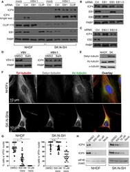
- Figure 1. Stable MTs Mediate EB-Independent HSV-1 Infection of SK-N-SHs (A) NHDFs or SK-N-SHs treated with non-targeting (ctrl), EB1, or CLIP-170 (CLIP) siRNAs were mock-infected or infected with HSV-1 at MOI 10 for 5 h and analyzed by WB. (B) SK-N-SHs treated with the indicated siRNAs were infected, as in (A). (C) SK-N-SHs treated with independent EB2 siRNAs (I or II) were infected, as in (A). (D) SK-N-SHs were treated with 100 nM BafA or DMSO and infected at MOI 10 with VSV for 4 h or HSV-1 for 5 h. (E) NHDFs or SK-N-SHs analyzed by WB using the indicated antibodies. (F) NHDFs or SK-N-SHs stained for tyrosinated (Tyr), detyrosinated (Detyr), and acetylated (Ac) tubulin. Nuclei were stained with Hoechst. (G) NHDFs or SK-N-SHs treated with 500 nM DMSO or 10 muM nocodazole were infected at MOI 20 with HSV-1 for 4 h in the presence of 1 mug/mL actinomycin D. Fixed cells were stained for VP5 and with Hoechst. Assessed for the accumulation of VP5 over 2 biological replicates were >= 165 NHDF or >= 190 SK-N-SH nuclei; error bars, SEMs; *p < 0.05, **p < 0.01, N.S., not significant; unpaired 2-tailed t test. (H) Cells treated as in (G) were infected at MOI 10 with HSV-1 for 5 h. All of the experiments represent >=3 replicates unless indicated. See also Figures S1 and S2 .

- Submitted by
- Invitrogen Antibodies (provider)
- Main image
- Experimental details
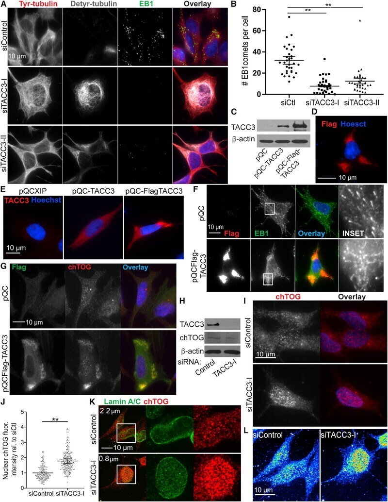
- Figure 3. TACC3 Regulates MT Dynamics and chTOG Localization (A) SK-N-SHs were treated with siRNAs and then fixed and stained for tyrosinated (Tyr) and detyrosinated (Detyr) tubulin, EB1, and Hoechst. Representative cells are shown. (B) Average number of EB1 comets per cell treated as in (A). Total number of EB1 comets in 30 cells per siRNA over 3 biological replicates; error bars, SEMs; **p < 0.01; unpaired 2-tailed t test. (C) SK-N-SHs were transduced with pQC, pQC-TACC3, or pQCXIP-FLAG-TACC3 (pQC-FLAG-TACC3) retroviral vectors and analyzed by WB. (D) SK-N-SHs transfected with pQC-FLAGTACC3 and fixed and stained for FLAG and with Hoechst. (E) SK-N-SHs transduced as in (C) were fixed and stained for TACC3 and Hoechst. (F) SK-N-SHs were transfected with pQC or pQC-FLAG-TACC3, fixed after 3 days, and stained for FLAG (red), EB1 (green), and with Hoechst. Enlarged image shows elongated EB1 staining patterns, indicative of reduced MT polymerization, in cells expressing FLAG-TACC3. (G) SK-N-SHs as in (F) were fixed and stained for FLAG (green) and chTOG (red) and with Hoechst. (H) SK-N-SHs treated with control or TACC3 siRNA and analyzed by WB. (I) siRNA-treated SK-N-SHs fixed and stained for chTOG (red) and with Hoechst. (J) Average corrected total fluorescence intensity of nuclear chTOG in SK-N-SHs treated with siRNAs; >= 255 cells per siRNA over 3 biological replicates; error bars, SEMs; **p < 0.01; unpaired 2-tailed t test. (K) SK-N-SHs were treated with siRNAs, fixed and sta