 Explore
 Validate
 Learn
Learn Western blot
 Gel shift
Antibody data
- Antibody Data
- Antigen structure
- References [0]
- Comments [0]
- Validations
- Western blot [3]
Submit
Validation data
Reference
Comment
Report error
- Product number
- PA5-19425 - Provider product page

- Provider
- Invitrogen Antibodies
- Product name
- Anti-Bub3 Polyclonal Antibody
- Antibody type
- Polyclonal
- Antigen
- Synthetic peptide
- Description
- This antibody is predicted to react with mouse, rat, Xenopus laevis and zebrafish based on sequence homology.
- Reactivity
- Human, Mouse
- Host
- Rabbit
- Isotype
- IgG
- Vial size
- 100 µg
- Concentration
- 0.5 mg/mL
- Storage
- Store at 4°C short term. For long term storage, store at -20°C, avoiding freeze/thaw cycles.
No comments: Submit comment
Supportive validation
- Submitted by
- Invitrogen Antibodies (provider)
- Main image
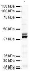
- Experimental details
- Western blot analysis of HeLa nuclear lysate using Product # PA5-19425, Bub3 primary antibody at a dilution of 1 µg/mL. Blot treated with a secondary HRP-conjugated Goat polyclonal anti-Rabbit antibody was used at a dilution of 1:10000.

- Submitted by
- Invitrogen Antibodies (provider)
- Main image
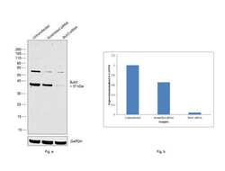
- Experimental details
- KD of Bub3 was achieved by transfecting HeLa with Bub3 specific siRNAs (Silencer® select Product # s17556, s2451). Western blot analysis (Fig. a) was performed using modified whole cell extracts (1% SDS) from the Bub3 KD cells (Lane 3), non-specific scrambled siRNA transfected cells (Lane 2) and untransfected cells (Lane 1). The blot was probed with Bub3 Polyclonal Antibody (Product # PA5-19425, 1:1000 dilution) and Goat anti-Rabbit IgG (H+L) Superclonal™ Secondary Antibody, HRP conjugate (Product # A27036, 1:4000 dilution). Densitometric analysis of this western blot is shown in histogram (Fig. b). Decrease in signal upon siRNA mediated knock down confirms that antibody is specific to Bub3. Certain uncharacterized bands were observed around ~55kDa..

- Submitted by
- Invitrogen Antibodies (provider)
- Main image
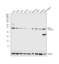
- Experimental details
- Western blot was performed using Anti-Bub3 Polyclonal Antibody (Product # PA5-19425) and 37 kDa band corresponding to Bub3 was observed across cell lines and tissue tested. Modified whole cell extracts (1% SDS) (30 µg lysate) of HeLa (Lane 1), A549 (Lane 2), A-431 (Lane 3), Hep G2 (Lane 4), PC-3 (Lane 5), HEK-293 (Lane 6), Jurkat (Lane 7), SK-BR-3 (Lane 8) and tissue extract of Mouse Spleen (Lane 9) were electrophoresed using Novex® NuPAGE® 4-12 % Bis-Tris gel (Product # NP0322BOX). Resolved proteins were then transferred onto a nitrocellulose membrane (Product # IB23001) by iBlot® 2 Dry Blotting System (Product # IB21001). The blot was probed with the primary antibody (1:1000 dilution) and detected by chemiluminescence Goat Anti-Rabbit IgG Secondary Antibody, HRP conjugate (Product # A27036, 1:4000 dilution) using the iBright FL 1000 (Product # A32752). Chemiluminescent detection was performed using Novex® ECL Chemiluminescent Substrate Reagent Kit (Product # WP20005). .
