 Explore
 Validate
 Learn
Learn Immunohistochemistry
Antibody data
- Antibody Data
- Antigen structure
- References [7]
- Comments [0]
- Validations
- Immunohistochemistry [1]
- Flow cytometry [2]
Submit
Validation data
Reference
Comment
Report error
- Product number
- MAB1621-100 - Provider product page

- Provider
- R&D Systems
- Product name
- Human DC-SIGN+DC-SIGNR Antibody
- Antibody type
- Monoclonal
- Description
- Protein A or G purified from hybridoma culture supernatant. Recognizes both human DC-SIGN and human DC-SIGNR on transfected cells. Does not react with parental mouse cells or irrelevant transfectants.
- Reactivity
- Human
- Host
- Mouse
- Conjugate
- Unconjugated
- Antigen sequence
Q9H2X3- Isotype
- IgG
- Antibody clone number
- 120612
- Vial size
- 100 ug
- Concentration
- LYOPH
- Storage
- Use a manual defrost freezer and avoid repeated freeze-thaw cycles. 12 months from date of receipt, -20 to -70 °C as supplied. 1 month, 2 to 8 °C under sterile conditions after reconstitution. 6 months, -20 to -70 °C under sterile conditions after reconstitution.
Submitted references Primary Human Placental Trophoblasts are Permissive for Zika Virus (ZIKV) Replication.
Uukuniemi Virus as a Tick-Borne Virus Model.
Binding of HIV-1 gp120 to DC-SIGN promotes ASK-1-dependent activation-induced apoptosis of human dendritic cells.
Vaccine protection by live, attenuated simian immunodeficiency virus in the absence of high-titer antibody responses and high-frequency cellular immune responses measurable in the periphery.
L-SIGN (CD209L) isoforms differently mediate trans-infection of hepatoma cells by hepatitis C virus pseudoparticles.
CCR5-, DC-SIGN-dependent endocytosis and delayed reverse transcription after human immunodeficiency virus type 1 infection in human astrocytes.
Rhesus macaque dendritic cells efficiently transmit primate lentiviruses independently of DC-SIGN.
Aagaard KM, Lahon A, Suter MA, Arya RP, Seferovic MD, Vogt MB, Hu M, Stossi F, Mancini MA, Harris RA, Kahr M, Eppes C, Rac M, Belfort MA, Park CS, Lacorazza D, Rico-Hesse R
Scientific reports 2017 Jan 27;7:41389
Scientific reports 2017 Jan 27;7:41389
Uukuniemi Virus as a Tick-Borne Virus Model.
Mazelier M, Rouxel RN, Zumstein M, Mancini R, Bell-Sakyi L, Lozach PY
Journal of virology 2016 Aug 1;90(15):6784-98
Journal of virology 2016 Aug 1;90(15):6784-98
Binding of HIV-1 gp120 to DC-SIGN promotes ASK-1-dependent activation-induced apoptosis of human dendritic cells.
Chen Y, Hwang SL, Chan VS, Chung NP, Wang SR, Li Z, Ma J, Lin CW, Hsieh YJ, Chang KP, Kung SS, Wu YC, Chu CW, Tai HT, Gao GF, Zheng B, Yokoyama KK, Austyn JM, Lin CL
PLoS pathogens 2013 Jan;9(1):e1003100
PLoS pathogens 2013 Jan;9(1):e1003100
Vaccine protection by live, attenuated simian immunodeficiency virus in the absence of high-titer antibody responses and high-frequency cellular immune responses measurable in the periphery.
Mansfield K, Lang SM, Gauduin MC, Sanford HB, Lifson JD, Johnson RP, Desrosiers RC
Journal of virology 2008 Apr;82(8):4135-48
Journal of virology 2008 Apr;82(8):4135-48
L-SIGN (CD209L) isoforms differently mediate trans-infection of hepatoma cells by hepatitis C virus pseudoparticles.
Falkowska E, Durso RJ, Gardner JP, Cormier EG, Arrigale RA, Ogawa RN, Donovan GP, Maddon PJ, Olson WC, Dragic T
The Journal of general virology 2006 Sep;87(Pt 9):2571-6
The Journal of general virology 2006 Sep;87(Pt 9):2571-6
CCR5-, DC-SIGN-dependent endocytosis and delayed reverse transcription after human immunodeficiency virus type 1 infection in human astrocytes.
Deiva K, Khiati A, Hery C, Salim H, Leclerc P, Horellou P, Tardieu M
AIDS research and human retroviruses 2006 Nov;22(11):1152-61
AIDS research and human retroviruses 2006 Nov;22(11):1152-61
Rhesus macaque dendritic cells efficiently transmit primate lentiviruses independently of DC-SIGN.
Wu L, Bashirova AA, Martin TD, Villamide L, Mehlhop E, Chertov AO, Unutmaz D, Pope M, Carrington M, KewalRamani VN
Proceedings of the National Academy of Sciences of the United States of America 2002 Feb 5;99(3):1568-73
Proceedings of the National Academy of Sciences of the United States of America 2002 Feb 5;99(3):1568-73
No comments: Submit comment
Supportive validation
- Submitted by
- R&D Systems (provider)
- Main image
- Experimental details
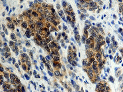
- DC-SIGN+DC-SIGNR in Human Lymphoma. DC-SIGN+DC-SIGNR was detected in immersion fixed paraffin-embedded sections of human lymphoma using 25 µg/mL Mouse Anti-Human DC-SIGN+ DC-SIGNR Monoclonal Antibody (Catalog # MAB1621) overnight at 4 °C. Tissue was stained with the Anti-Mouse HRP-DAB Cell & Tissue Staining Kit (brown; Catalog # CTS002) and counter-stained with hematoxylin (blue). View our protocol for Chromogenic IHC Staining of Paraffin-embedded Tissue Sections.

Supportive validation
- Submitted by
- R&D Systems (provider)
- Main image

- Experimental details
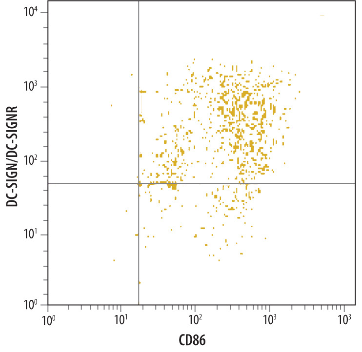
- Detection of DC-SIGN+DC-SIGNR in Human Monocyte Derived Dendritic Cells by Flow Cytometry. Human monocyte derived dendritic cells were stained with Mouse Anti-Human DC-SIGN+ DC-SIGNR Monoclonal Antibody (Catalog # MAB1621), followed by PE-conjugated anti-mouse secondary antibody (Catalog # F0102B) and Human B7-2/CD86 Fluorescein-conjugated Monoclonal Antibody (Catalog # FAB141F).Quadrant markers were set based on isotype control antibody staining (Catalog # MAB003).
- Submitted by
- R&D Systems (provider)
- Main image
- Experimental details
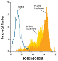
- Detection of DC-SIGN+DC-SIGNR in Human DC-SIGN or DC-SIGNR Transfected 3T3 Mouse Cell Line by Flow Cytometry. Human DC-SIGN and DC-SIGNR transfected 3T3 mouse embryonic fibroblast cell line were stained with Mouse Anti-Human DC-SIGN+ DC-SIGNR Monoclonal Antibody (Catalog # MAB1621, filled histograms) or isotype control antibody (Catalog # MAB003, open histogram), followed by Phycoerythrin-conjugated Anti-Mouse IgG F(ab')2 Secondary Antibody (Catalog # F0102B).
