Explore
 Validate
 Learn
Learn Western blot
Antibody data
- Antibody Data
- Antigen structure
- References [0]
- Comments [0]
- Validations
- Western blot [2]
- Flow cytometry [1]
Submit
Validation data
Reference
Comment
Report error
- Product number
- 10-10020-25 - Provider product page

- Provider
- ABEOMICS Inc.
- Product name
- Anti-Dc-Sign/CD 209 Antibody
- Antibody type
- Monoclonal
- Description
- Dendritic cell-specific intercellular adhesion molecule-3-grabbing non-integrin (DC-SIGN) is a tetrameric C-type (calcium-dependent) lectin that binds, through its C-terminal carbohydrate recognition domain, high mannose N-linked glycans present on the surface of several viral glycoproteins such as human immunodeficiency virus (HIV) gp120 and hepatitis C virus (HCV) E2. It facilitates DC-specific delivery of Ag. This is accomplished by conjugating Ag to receptor-specific Ab or carbohydrate ligands that bind to its carbohydrate recognition domain. In humans, DC-SIGN expression is restricted to DCs and certain types of macrophages. DC-SIGN is involved in the innate immune system and recognizes numerous evolutionarily divergent pathogens, including viruses, bacteria, fungi, and parasites. After binding, these pathogens are internalized and pathogen-derived antigens are presented via MHC class I and II molecules to CD8+ and CD4+ T cells, respectively. DC-SIGN represents a promising CLR for targeted vaccine delivery.
- Reactivity
- Human
- Host
- Mouse
- Conjugate
- Unconjugated
- Antigen sequence
A recombinant protein fragment of D
C-Sign protein was used as the immu
nogen for this antibody.- Isotype
- IgG
- Antibody clone number
- ABM47B7
- Vial size
- 100 µg
- Concentration
- 0.5 mg/ml
- Storage
- Store the antibody at 4°C, stable for 6 months. For long-term storage, store at -20°C. Avoid repeat freez thawing
No comments: Submit comment
Supportive validation
- Submitted by
- ABEOMICS Inc. (provider)
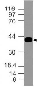
- Main image

- Experimental details
- Expression analysis of Dc-Sign. Anti-Dc-Sign antibody (Clone: ABM47B7) was tested at 0.1 µg/ml on partial length recombinant protein.
- Protocol
- Protocol
- Submitted by
- ABEOMICS Inc. (provider)
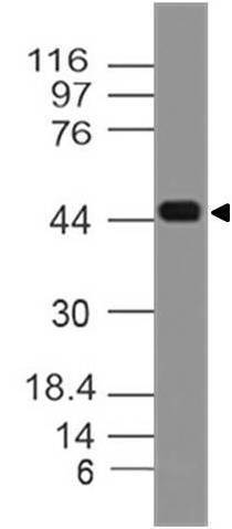
- Main image

- Experimental details
- Expression analysis of Dc-Sign. Anti-Dc-Sign antibody (Clone: ABM47B7) was tested at 0.1 µg/ml on human Liver lysate.
- Protocol
- Protocol
Supportive validation
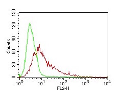
- Submitted by
- ABEOMICS Inc. (provider)
- Main image
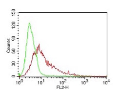
- Experimental details
- Cell Surface FLOW analysis of Dc-Sign antibody (10-10020) in 293HEK-Dc-sign stable cell line using 0.5 µg/ 10^6 cells. Green represents isotype control, red represents anti- Dc-Sign antibody. Goat anti-mouse PE conjugates was used as secondary antibody.
- Protocol
- Protocol