Explore
 Validate
 Learn
Learn Flow cytometry
Antibody data
- Antibody Data
- Antigen structure
- References [8]
- Comments [0]
- Validations
- Flow cytometry [1]
- Other assay [3]
Submit
Validation data
Reference
Comment
Report error
- Product number
- 14-2099-37 - Provider product page

- Provider
- Invitrogen Antibodies
- Product name
- CD209 (DC-SIGN) Monoclonal Antibody (eB-h209), eBioscience™
- Antibody type
- Monoclonal
- Antigen
- Other
- Description
- Description: The eB-h209 monoclonal antibody reacts with human CD209, also known as DC-SIGN, a 44 kDa type II transmembrane protein. DC-SIGN contains a C-type lectin binding domain and binds ICAM-3, ICAM-2, and HIV virus. Human dendritic cells preferentially express DC-SIGN. It has been postulated that DC-SIGN serves as a receptor for capture, trafficking, and transmission of HIV to T cells and supports primary immune response. eB-h209 was developed against a C-terminal peptide of human DC-SIGN.
- Antibody clone number
- eB-h209
- Concentration
- 0.5 mg/mL
Submitted references Different Induction of PD-L1 (CD274) and PD-1 (CD279) Expression in THP-1-Differentiated Types 1 and 2 Macrophages.
Activation of Human Vδ2(+) γδ T Cells by Staphylococcus aureus Promotes Enhanced Anti-Staphylococcal Adaptive Immunity.
Porphyromonas gingivalis evasion of autophagy and intracellular killing by human myeloid dendritic cells involves DC-SIGN-TLR2 crosstalk.
Human Blood-Circulating Basophils Capture HIV-1 and Mediate Viral trans-Infection of CD4+ T Cells.
Dectin-1/TLR2 and NOD2 agonists render dendritic cells susceptible to infection by X4-using HIV-1 and promote cis-infection of CD4(+) T cells.
A consensus surface activation marker signature is partially dependent on human immunodeficiency virus type 1 Nef expression within productively infected macrophages.
CD137 ligand signaling induces human monocyte to dendritic cell differentiation.
Phenotypic modulation of the stromal reticular network in normal and neoplastic lymph nodes: tissue transglutaminase reveals coordinate regulation of multiple cell types.
Lai CY, Tseng PC, Chen CL, Satria RD, Wang YT, Lin CF
Journal of inflammation research 2021;14:5241-5249
Journal of inflammation research 2021;14:5241-5249
Activation of Human Vδ2(+) γδ T Cells by Staphylococcus aureus Promotes Enhanced Anti-Staphylococcal Adaptive Immunity.
Cooper AJR, Lalor SJ, McLoughlin RM
Journal of immunology (Baltimore, Md. : 1950) 2020 Aug 15;205(4):1039-1049
Journal of immunology (Baltimore, Md. : 1950) 2020 Aug 15;205(4):1039-1049
Porphyromonas gingivalis evasion of autophagy and intracellular killing by human myeloid dendritic cells involves DC-SIGN-TLR2 crosstalk.
El-Awady AR, Miles B, Scisci E, Kurago ZB, Palani CD, Arce RM, Waller JL, Genco CA, Slocum C, Manning M, Schoenlein PV, Cutler CW
PLoS pathogens 2015 Feb;10(2):e1004647
PLoS pathogens 2015 Feb;10(2):e1004647
Human Blood-Circulating Basophils Capture HIV-1 and Mediate Viral trans-Infection of CD4+ T Cells.
Jiang AP, Jiang JF, Guo MG, Jin YM, Li YY, Wang JH
Journal of virology 2015 Aug;89(15):8050-62
Journal of virology 2015 Aug;89(15):8050-62
Dectin-1/TLR2 and NOD2 agonists render dendritic cells susceptible to infection by X4-using HIV-1 and promote cis-infection of CD4(+) T cells.
Côté SC, Plante A, Tardif MR, Tremblay MJ
PloS one 2013;8(7):e67735
PloS one 2013;8(7):e67735
A consensus surface activation marker signature is partially dependent on human immunodeficiency virus type 1 Nef expression within productively infected macrophages.
Babu R, Brown A
Retrovirology 2013 Dec 16;10:155
Retrovirology 2013 Dec 16;10:155
CD137 ligand signaling induces human monocyte to dendritic cell differentiation.
Kwajah M M S, Schwarz H
European journal of immunology 2010 Jul;40(7):1938-49
European journal of immunology 2010 Jul;40(7):1938-49
Phenotypic modulation of the stromal reticular network in normal and neoplastic lymph nodes: tissue transglutaminase reveals coordinate regulation of multiple cell types.
Thomazy VA, Vega F, Medeiros LJ, Davies PJ, Jones D
The American journal of pathology 2003 Jul;163(1):165-74
The American journal of pathology 2003 Jul;163(1):165-74
No comments: Submit comment
Supportive validation
- Submitted by
- Invitrogen Antibodies (provider)
- Main image
- Experimental details
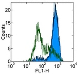
- Staining of human monocyte-derived immature dendritic cells with 0.5 µg of Rat IgG2a K Isotype Control Purified (Product # 14-4321-82) (open histogram) or 0.5 µg of Anti-Human CD209 (DC-SIGN) Purified (filled histogram) followed by Anti-Rat IgG FITC (Product # 11-4811-85). Cells in the large scatter population were used for analysis.

Supportive validation
- Submitted by
- Invitrogen Antibodies (provider)
- Main image

- Experimental details
- NULL
- Submitted by
- Invitrogen Antibodies (provider)
- Main image
- Experimental details
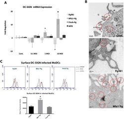
- Fig 2 Mfa1 + Pg up-regulate the expression of DC-SIGN in human MoDCs. A) DC-SIGN mRNA expression in P. gingivalis -infected MoDCs at 0.1, 1 and 10 MOIs. The figure shows the gene expression after 12 hours of Pg381 and mutant strains infections. The target gene (DC-SIGN) was normalized using the endogenous control GAPDH (DeltaCt) and fold regulations were calculated using 2 -(DeltaDeltaCt) method. The statistical analysis was performed using the t-test , which accounts for the clustering of infected and un-infected controls within 3 different experiments (* p

- Submitted by
- Invitrogen Antibodies (provider)
- Main image
- Experimental details
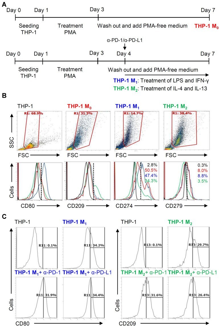
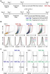
- Figure 5 Different expression of PD-L1 (CD274) and PD-1 (CD279) in THP-1-differentiated macrophages. ( A ) In PMA-stimulated THP-1-differentiated M 0 macrophages, cells were then treated with LPS (1 mug/mL)/IFN-gamma (10 ng/mL) and IL-4 (25 ng/mL)/IL-13 (25 ng/mL) for polarization of M 1 and M 2 , respectively, in the absence and presence of neutralizing antibodies (5 mug/mL) against CD274 (alpha-PD-L1) and CD279 (alpha-PD-1) according to the experimental design. ( B ) For immunostaining, cells were stained with CD80 and CD209 for dissecting M 1 and M 2 , respectively. Immunostaining followed by flow cytometric histogram analysis showed the expression of CD274 and CD279 in these cells. ( C ) Furthermore, the expression of CD80 and CD209 in M 1 and M 2 without or with the blockade of CD274 and CD279 were shown. For all flow cytometric analysis, representative data were selectively obtained from three individual experiments, and the percentage of positive cells is shown. THP-1 (black); THP-1 M 0 (red); THP-1 M 1 (blue); THP-1 M 2 (green).