Explore
 Validate
 Learn
Learn Western blot
 ELISA
ELISA Immunocytochemistry
Antibody data
- Antibody Data
- Antigen structure
- References [1]
- Comments [0]
- Validations
- Immunocytochemistry [2]
- Immunohistochemistry [7]
- Flow cytometry [2]
- Other assay [1]
Submit
Validation data
Reference
Comment
Report error
- Product number
- PA5-79529 - Provider product page

- Provider
- Invitrogen Antibodies
- Product name
- CD49e (Integrin alpha 5) Polyclonal Antibody
- Antibody type
- Polyclonal
- Antigen
- Recombinant full-length protein
- Description
- Reconstitute with 0.2 mL of distilled water to yield a concentration of 500 µg/mL. Positive Control - WB: human A549 whole cell lysates, human Hela whole cell lysates, human placenta tissue lysates, rat brain tissue lysates, rat PC-12 whole cell lysates, mouse brain tissue lysates. IHC: human placenta tissue, rat bladder tissue .
- Reactivity
- Human, Mouse, Rat
- Host
- Rabbit
- Isotype
- IgG
- Vial size
- 100 μg
- Concentration
- 500 μg/mL
- Storage
- -20°C
Submitted references Cell surface integrin α5ß1 clustering negatively regulates receptor tyrosine kinase signaling in colorectal cancer cells via glycogen synthase kinase 3.
Starchenko A, Graves-Deal R, Brubaker D, Li C, Yang Y, Singh B, Coffey RJ, Lauffenburger DA
Integrative biology : quantitative biosciences from nano to macro 2021 Jun 15;13(6):153-166
Integrative biology : quantitative biosciences from nano to macro 2021 Jun 15;13(6):153-166
No comments: Submit comment
Supportive validation
- Submitted by
- Invitrogen Antibodies (provider)
- Main image
- Experimental details
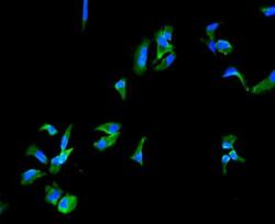
- Immunocytochemistry analysis of Integrin alpha 5 using anti-Integrin alpha 5 antibody (Product # PA5-79529) . Integrin alpha 5 was detected in a section of HeLa cells. Enzyme antigen retrieval was performed using IHC enzyme antigen retrieval reagent for 15 mins. The cells were blocked with 10% goat serum and then incubated with 2μg/mL rabbit anti-Integrin alpha 5 antibody (Product # PA5-79529) overnight at 4°C. DyLight®488 Conjugated Goat Anti-Rabbit IgG was used as secondary antibody at 1:100 dilution and incubated for 30 minutes at 37°C. The section was counterstained with DAPI. Visualize using a fluorescence microscope and filter sets appropriate for the label used.

- Submitted by
- Invitrogen Antibodies (provider)
- Main image

- Experimental details
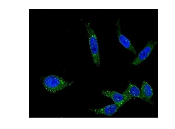
- Immunocytochemistry analysis of Integrin alpha 5 using anti-Integrin alpha 5 antibody (Product # PA5-79529) . Integrin alpha 5 was detected in a section of U2OS cells. Enzyme antigen retrieval was performed using IHC enzyme antigen retrieval reagent for 15 mins. The cells were blocked with 10% goat serum and then incubated with 2μg/mL rabbit anti-Integrin alpha 5 antibody (Product # PA5-79529) overnight at 4°C. DyLight®488 Conjugated Goat Anti-Rabbit IgG was used as secondary antibody at 1:100 dilution and incubated for 30 minutes at 37°C. The section was counterstained with DAPI. Visualize using a fluorescence microscope and filter sets appropriate for the label used.
Supportive validation
- Submitted by
- Invitrogen Antibodies (provider)
- Main image

- Experimental details
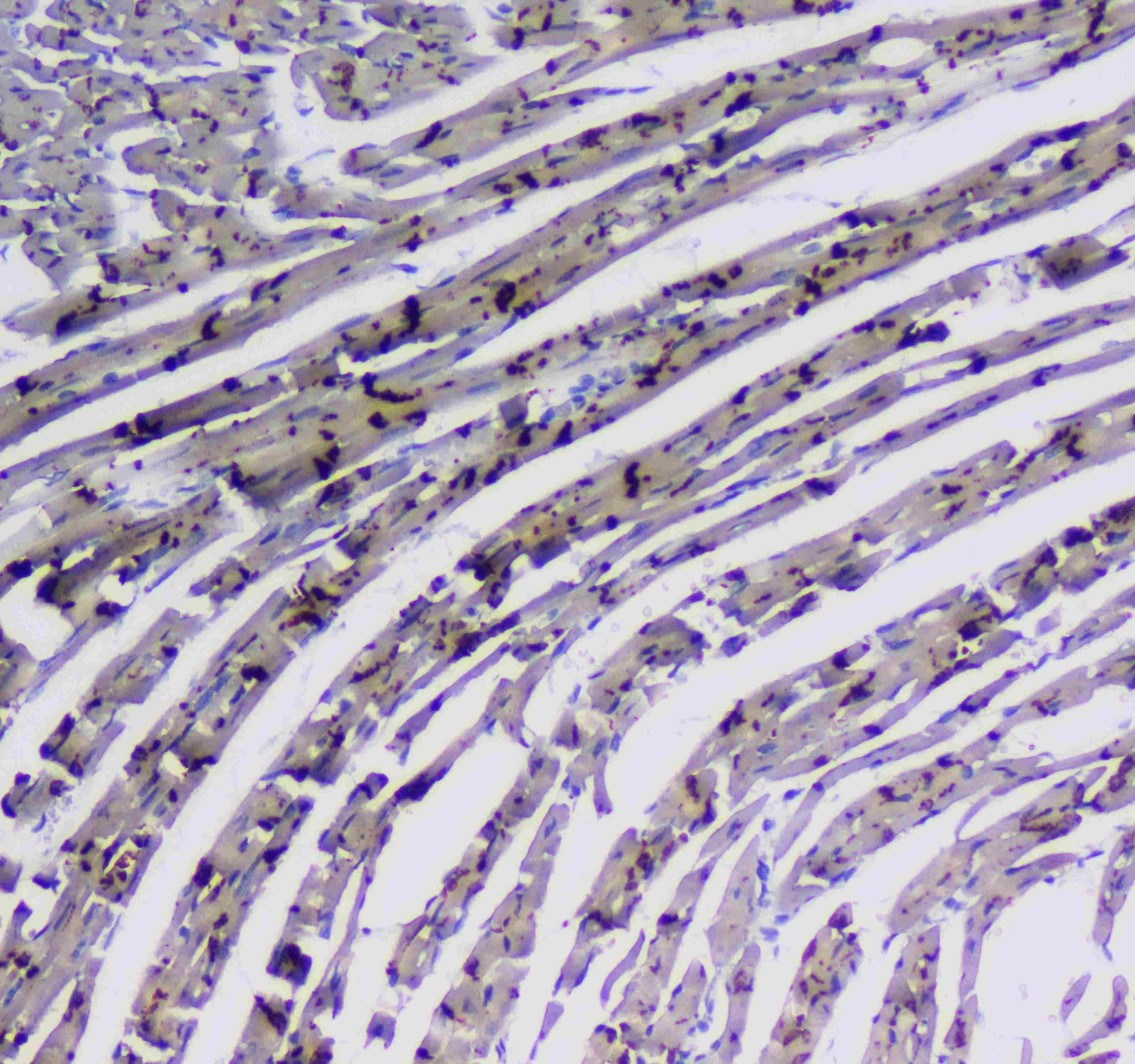
- Immunohistochemistry analysis of CD49e (Integrin alpha 5) on paraffin-embedded rat cardiac muscle tissue. Antigen retrieval was performed using citrate buffer (pH6, epitope retrieval solution) for 20 mins. Sample was blocked using 10% goat serum, incubated with CD49e (Integrin alpha 5) polyclonal antibody (Product# PA5-79529) with a dilution of 1 µg/mL (overnight at 4°C), and followed by biotinylated goat anti-rabbit IgG (30 minutes at 37°C). Development was performed using Streptavidin-Biotin-Complex (SABC) with DAB chromogen method.
- Submitted by
- Invitrogen Antibodies (provider)
- Main image
- Experimental details
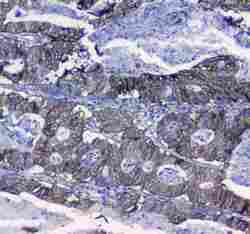
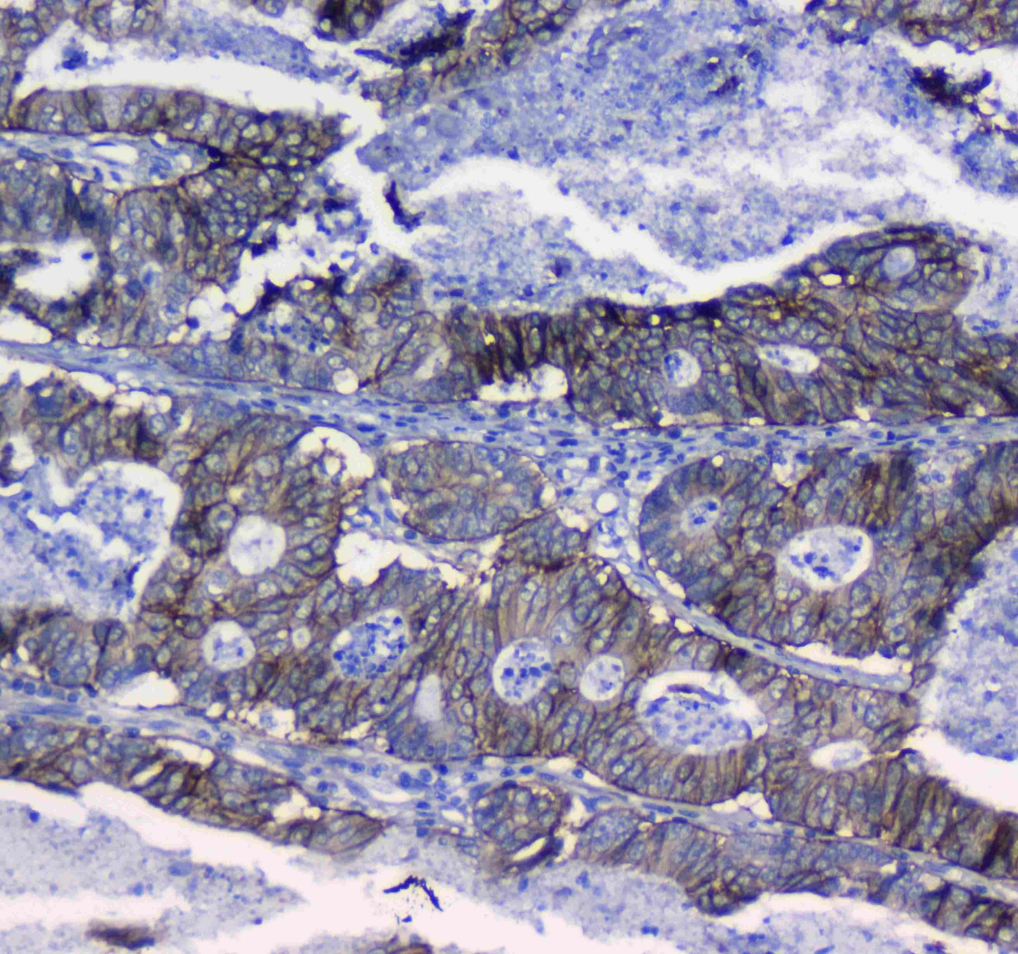
- Immunohistochemistry analysis of CD49e (Integrin alpha 5) on paraffin-embedded human colon cancer tissue. Antigen retrieval was performed using citrate buffer (pH6, epitope retrieval solution) for 20 mins. Sample was blocked using 10% goat serum, incubated with CD49e (Integrin alpha 5) polyclonal antibody (Product# PA5-79529) with a dilution of 1 µg/mL (overnight at 4°C), and followed by biotinylated goat anti-rabbit IgG (30 minutes at 37°C). Development was performed using Streptavidin-Biotin-Complex (SABC) with DAB chromogen method.
- Submitted by
- Invitrogen Antibodies (provider)
- Main image
- Experimental details
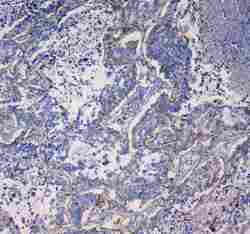
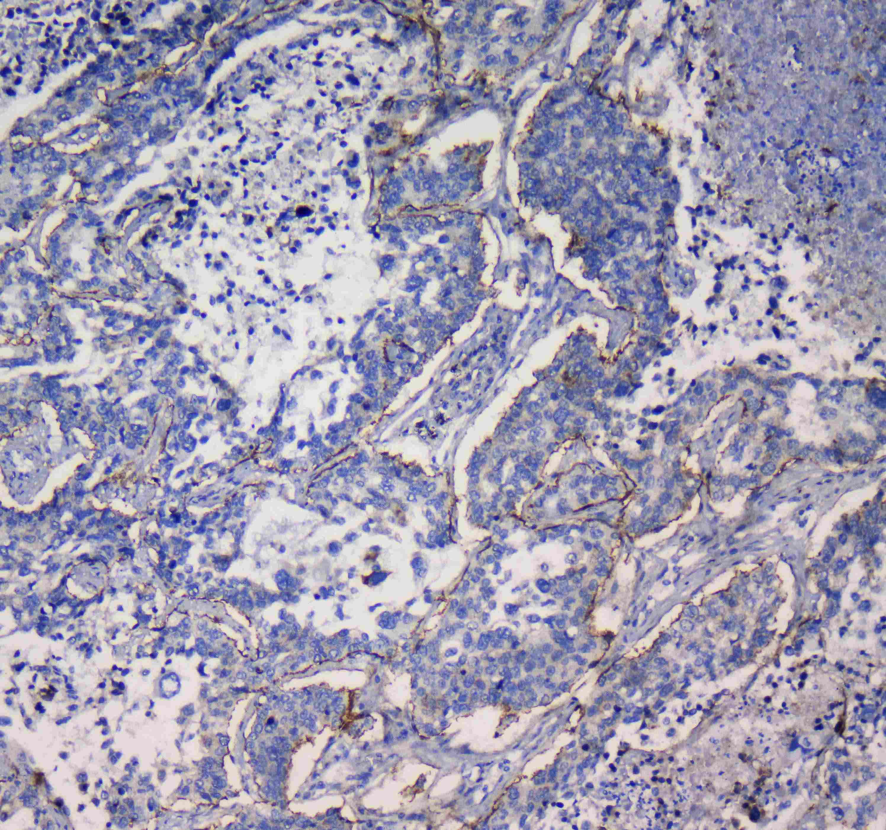
- Immunohistochemistry analysis of CD49e (Integrin alpha 5) on paraffin-embedded human lung cancer tissue. Antigen retrieval was performed using citrate buffer (pH6, epitope retrieval solution) for 20 mins. Sample was blocked using 10% goat serum, incubated with CD49e (Integrin alpha 5) polyclonal antibody (Product# PA5-79529) with a dilution of 1 µg/mL (overnight at 4°C), and followed by biotinylated goat anti-rabbit IgG (30 minutes at 37°C). Development was performed using Streptavidin-Biotin-Complex (SABC) with DAB chromogen method.
- Submitted by
- Invitrogen Antibodies (provider)
- Main image

- Experimental details
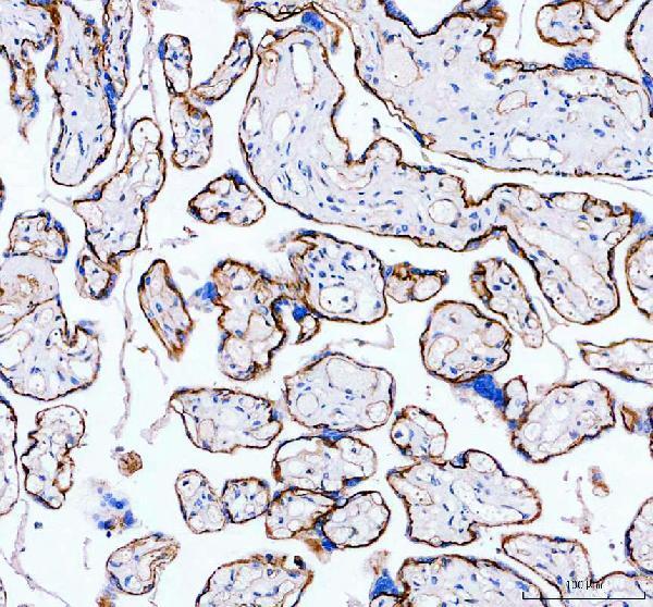
- Immunohistochemistry (Paraffin) analysis of CD49e (Integrin alpha 5) in paraffin-embedded section of human placenta tissue using CD49e (Integrin alpha 5) Polyclonal Antibody (Product # PA5-79529). Heat mediated antigen retrieval was performed in EDTA buffer (pH 8.0, epitope retrieval solution). The tissue section was blocked with 10% goat serum. The tissue section was then incubated with the primary antibody at a 2 µg/mL dilution overnight at 4°C. Peroxidase conjugated goat anti-rabbit IgG was used as secondary antibody and incubated for 30 minutes at 37°C. The tissue section was developed using HRP Conjugated Rabbit IgG Super Vision Assay Kit with DAB as the chromogen.
- Submitted by
- Invitrogen Antibodies (provider)
- Main image

- Experimental details
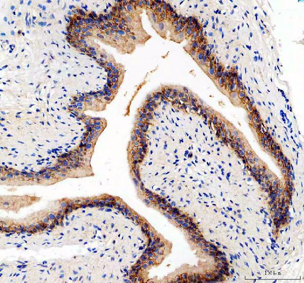
- Immunohistochemistry (Paraffin) analysis of CD49e (Integrin alpha 5) in paraffin-embedded section of rat bladder tissue using CD49e (Integrin alpha 5) Polyclonal Antibody (Product # PA5-79529). Heat mediated antigen retrieval was performed in EDTA buffer (pH 8.0, epitope retrieval solution). The tissue section was blocked with 10% goat serum. The tissue section was then incubated with the primary antibody at a 2 µg/mL dilution overnight at 4°C. Peroxidase conjugated goat anti-rabbit IgG was used as secondary antibody and incubated for 30 minutes at 37°C. The tissue section was developed using HRP Conjugated Rabbit IgG Super Vision Assay Kit with DAB as the chromogen.
- Submitted by
- Invitrogen Antibodies (provider)
- Main image
- Experimental details
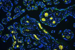
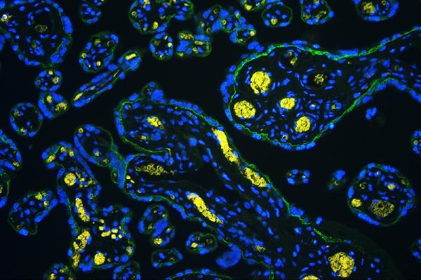
- Immunohistochemical analysis of Integrin alpha 5 in paraffin-embedded section of human placenta tissues. Heat mediated antigen retrieval was performed in citrate buffer (pH6, epitope retrieval solution ) for 20 mins. The tissue section was blocked with 10% goat serum. The tissue section was then incubated with 1μg/mL rabbit anti-Integrin alpha 5 antibody (Product # PA5-79529) overnight at 4°C. DyLight®488 Conjugated Goat Anti-Rabbit IgG was used as secondary antibody at 1:100 dilution and incubated for 30 minutes at 37°C. The section was counterstained with DAPI. Visualize using a fluorescence microscope and filter sets appropriate for the label used.
- Submitted by
- Invitrogen Antibodies (provider)
- Main image
- Experimental details
- Immunohistochemical analysis of Integrin alpha 5 in paraffin-embedded section of human colon cancer tissues. Heat mediated antigen retrieval was performed in citrate buffer (pH6, epitope retrieval solution ) for 20 mins. The tissue section was blocked with 10% goat serum. The tissue section was then incubated with 1μg/mL rabbit anti-Integrin alpha 5 antibody (Product # PA5-79529) overnight at 4°C. DyLight®488 Conjugated Goat Anti-Rabbit IgG was used as secondary antibody at 1:100 dilution and incubated for 30 minutes at 37°C. The section was counterstained with DAPI. Visualize using a fluorescence microscope and filter sets appropriate for the label used.
Supportive validation
- Submitted by
- Invitrogen Antibodies (provider)
- Main image
- Experimental details
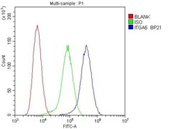
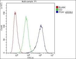
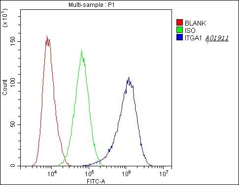
- Flow Cytometry of CD49e (Integrin alpha 5) in HT-1080 cells (blue line), isotype control rabbit IgG (green line) and unlabeled (red line). Samples were blocked with 10% goat serum, incubated with CD49e (Integrin alpha 5) Polyclonal Antibody (Product # PA5-79529) at a dilution of 1 μg (per 1x10^6 cells), followed by DyLight®488 conjugated goat anti-rabbit IgG (for 30 minutes at 20°C) using 5-10 μg (per 1x10^6 cells) dilution.
- Submitted by
- Invitrogen Antibodies (provider)
- Main image
- Experimental details
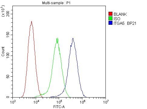
- Flow Cytometry of CD49e (Integrin alpha 5) in A431 cells (blue line), isotype control rabbit IgG (green line) and unlabeled (red line). Samples were blocked with 10% goat serum, incubated with CD49e (Integrin alpha 5) Polyclonal Antibody (Product # PA5-79529) at a dilution of 1 μg (per 1x10^6 cells), followed by DyLight®488 conjugated goat anti-rabbit IgG (for 30 minutes at 20°C) using 5-10 μg (per 1x10^6 cells) dilution.
Supportive validation
- Submitted by
- Invitrogen Antibodies (provider)
- Main image

- Experimental details
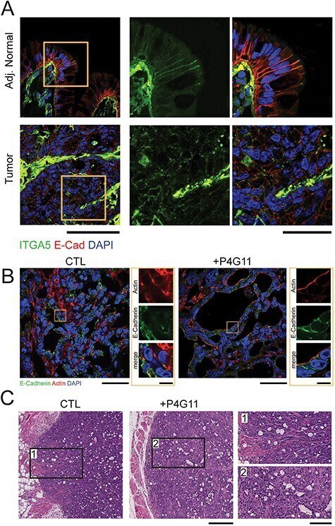
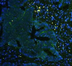
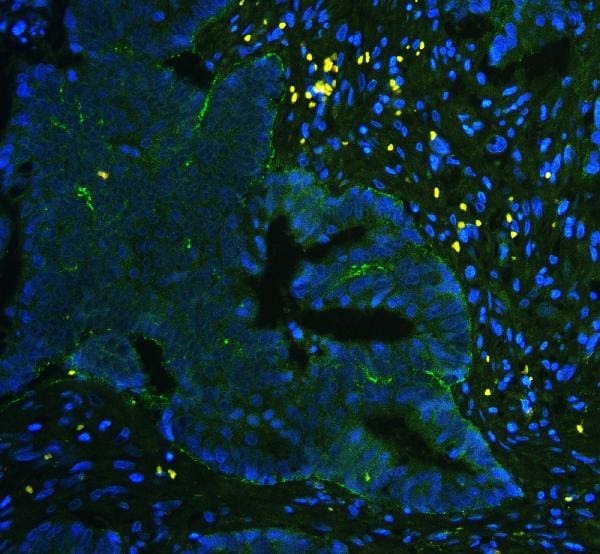
- Figure 1 Presence of Integrin alpha5ss1 at lateral cell-cell junctions correlates with increased epithelial organization in vivo. (A) FFPE sections from human colorectal cancer tumors and adjacent normal regions were stained for integrin alpha5 (green), E-Cadherin (red), and DAPI (blue) (B-C) HCA-7-derived SC cells were injected subcutaneously into nude mice and allowed to establish palpable tumors (1 week). Mice were subsequently given P4G11 or vehicle via intraperitoneal injection for 21 days (B) Representative confocal images of SC tumor sections stained with antibody against E-Cadherin (green), Phalloidin (red) and DAPI (blue). Scale bar = 50um. Inset scale bar = 10um. Note appearance of basolateral antimouse secondary antibody binding in P4G11-treated tumors. (C) Representative histology of control and P4G11-treated tumors, scale bar = 500um, inset scale bar = 50uM.