Explore
 Validate
 Learn
Learn Western blot
 Immunohistochemistry
Antibody data
- Antibody Data
- Antigen structure
- References [4]
- Comments [0]
- Validations
- Immunohistochemistry [1]
Submit
Validation data
Reference
Comment
Report error
- Product number
- HPA002642 - Provider product page

- Provider
- Atlas Antibodies
- Proper citation
- Atlas Antibodies Cat#HPA002642, RRID:AB_1078469
- Product name
- Anti-ITGA5
- Antibody type
- Polyclonal
- Description
- Polyclonal Antibody against Human ITGA5, Gene description: integrin, alpha 5 (fibronectin receptor, alpha polypeptide), Alternative Gene Names: CD49e, FNRA, Validated applications: IHC, WB, Uniprot ID: P08648, Storage: Store at +4°C for short term storage. Long time storage is recommended at -20°C.
- Reactivity
- Human, Mouse
- Host
- Rabbit
- Conjugate
- Unconjugated
- Isotype
- IgG
- Vial size
- 100 µl
- Concentration
- 0.2 mg/ml
- Storage
- Store at +4°C for short term storage. Long time storage is recommended at -20°C.
- Handling
- The antibody solution should be gently mixed before use.
Submitted references ITGA5 inhibition in pancreatic stellate cells attenuates desmoplasia and potentiates efficacy of chemotherapy in pancreatic cancer
Integrin α5 subunit is required for the tumor supportive role of fibroblasts in colorectal adenocarcinoma and serves as a potential stroma prognostic marker
Single-Cell Transcriptomic Analysis of Primary and Metastatic Tumor Ecosystems in Head and Neck Cancer
Survivin promotion of melanoma metastasis requires upregulation of α 5 integrin
Kuninty P, Bansal R, De Geus S, Mardhian D, Schnittert J, van Baarlen J, Storm G, Bijlsma M, van Laarhoven H, Metselaar J, Kuppen P, Vahrmeijer A, Östman A, Sier C, Prakash J
Science Advances 2019;5(9)
Science Advances 2019;5(9)
Integrin α5 subunit is required for the tumor supportive role of fibroblasts in colorectal adenocarcinoma and serves as a potential stroma prognostic marker
Lu L, Xie R, Wei R, Cai C, Bi D, Yin D, Liu H, Zheng J, Zhang Y, Song F, Gao Y, Tan L, Wei Q, Qin H
Molecular Oncology 2019;13(12):2697-2714
Molecular Oncology 2019;13(12):2697-2714
Single-Cell Transcriptomic Analysis of Primary and Metastatic Tumor Ecosystems in Head and Neck Cancer
Puram S, Tirosh I, Parikh A, Patel A, Yizhak K, Gillespie S, Rodman C, Luo C, Mroz E, Emerick K, Deschler D, Varvares M, Mylvaganam R, Rozenblatt-Rosen O, Rocco J, Faquin W, Lin D, Regev A, Bernstein B
Cell 2017;171(7):1611-1624.e24
Cell 2017;171(7):1611-1624.e24
Survivin promotion of melanoma metastasis requires upregulation of α 5 integrin
McKenzie J, Liu T, Jung J, Jones B, Ekiz H, Welm A, Grossman D
Carcinogenesis 2013;34(9):2137-2144
Carcinogenesis 2013;34(9):2137-2144
No comments: Submit comment
Supportive validation
- Submitted by
- Atlas Antibodies (provider)
- Enhanced method
- Orthogonal validation
- Main image
- Experimental details
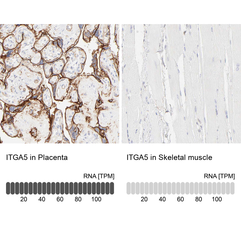
- Immunohistochemistry analysis in human placenta and skeletal muscle tissues using HPA002642 antibody. Corresponding ITGA5 RNA-seq data are presented for the same tissues.
- Sample type
- Human
- Protocol
- Protocol