Explore
 Validate
 Learn
Learn Western blot
Antibody data
- Antibody Data
- Antigen structure
- References [7]
- Comments [0]
- Validations
- Western blot [11]
- Immunohistochemistry [4]
- Other assay [5]
Submit
Validation data
Reference
Comment
Report error
- Product number
- MA1-91637 - Provider product page

- Provider
- Invitrogen Antibodies
- Product name
- Active/Pro-Caspase 3 Monoclonal Antibody (31A1067)
- Antibody type
- Monoclonal
- Antigen
- Recombinant full-length protein
- Description
- Predicted molecular weight: 31kDa.
- Antibody clone number
- 31A1067
- Concentration
- 1 mg/mL
Submitted references Aging lens epithelium is susceptible to ferroptosis.
Pristimerin inhibits neuronal inflammation and protects cognitive function in mice with sepsis-induced brain injuries by regulating PI3K/Akt signalling.
Mitotic Activation Around Wound Edges and Epithelialization Repair in UVB-Induced Capsular Cataracts.
β-arrestin 2 Is a Prognostic Factor for Survival of Ovarian Cancer Patients Upregulating Cell Proliferation.
Therapeutic effect of Resveratrol in the treatment of osteoarthritis via the MALAT1/miR-9/NF-κB signaling pathway.
Estrogen Modulates Specific Life and Death Signals Induced by LH and hCG in Human Primary Granulosa Cells In Vitro.
Esculetin induces antiproliferative and apoptotic response in pancreatic cancer cells by directly binding to KEAP1.
Wei Z, Hao C, Huangfu J, Srinivasagan R, Zhang X, Fan X
Free radical biology & medicine 2021 May 1;167:94-108
Free radical biology & medicine 2021 May 1;167:94-108
Pristimerin inhibits neuronal inflammation and protects cognitive function in mice with sepsis-induced brain injuries by regulating PI3K/Akt signalling.
Xue W, Li Y, Zhang M
Pharmaceutical biology 2021 Dec;59(1):1351-1358
Pharmaceutical biology 2021 Dec;59(1):1351-1358
Mitotic Activation Around Wound Edges and Epithelialization Repair in UVB-Induced Capsular Cataracts.
Wei Z, Hao C, Srinivasagan R, Wu H, Chen JK, Fan X
Investigative ophthalmology & visual science 2021 Dec 1;62(15):29
Investigative ophthalmology & visual science 2021 Dec 1;62(15):29
β-arrestin 2 Is a Prognostic Factor for Survival of Ovarian Cancer Patients Upregulating Cell Proliferation.
Czogalla B, Partenheimer A, Jeschke U, von Schönfeldt V, Mayr D, Mahner S, Burges A, Simoni M, Melli B, Benevelli R, Bertini S, Casarini L, Trillsch F
Frontiers in endocrinology 2020;11:554733
Frontiers in endocrinology 2020;11:554733
Therapeutic effect of Resveratrol in the treatment of osteoarthritis via the MALAT1/miR-9/NF-κB signaling pathway.
Zhang G, Zhang H, You W, Tang X, Li X, Gong Z
Experimental and therapeutic medicine 2020 Mar;19(3):2343-2352
Experimental and therapeutic medicine 2020 Mar;19(3):2343-2352
Estrogen Modulates Specific Life and Death Signals Induced by LH and hCG in Human Primary Granulosa Cells In Vitro.
Casarini L, Riccetti L, De Pascali F, Gilioli L, Marino M, Vecchi E, Morini D, Nicoli A, La Sala GB, Simoni M
International journal of molecular sciences 2017 Apr 28;18(5)
International journal of molecular sciences 2017 Apr 28;18(5)
Esculetin induces antiproliferative and apoptotic response in pancreatic cancer cells by directly binding to KEAP1.
Arora R, Sawney S, Saini V, Steffi C, Tiwari M, Saluja D
Molecular cancer 2016 Oct 18;15(1):64
Molecular cancer 2016 Oct 18;15(1):64
No comments: Submit comment
Supportive validation
- Submitted by
- Invitrogen Antibodies (provider)
- Main image

- Experimental details
- Western blot analysis of Caspase 3 active & pro in human A) brain, B) heart, C) intestine, D) kidney, E) liver, F) lung, G) muscle, H) stomach, I) spleen, J) ovary, and K) testis using a Caspase 3 active & pro monoclonal antibody (Product # MA1-91637) at a dilution of 5 µg/mL.

- Submitted by
- Invitrogen Antibodies (provider)
- Main image

- Experimental details
- Western Blot analysis of Caspase 3 (active/pro) was performed by loading HeLa cell lysate. Proteins were transferred to a membrane and probed with a Caspase 3 (active/pro) Monoclonal Antibody (31A1067) (Product # MA1-91637).

- Submitted by
- Invitrogen Antibodies (provider)
- Main image

- Experimental details
- Western Blot analysis of Caspase 3 (active/pro) was performed by loading human (A) brain, (B) heart, (C) intestine, (D) kidney, (E) liver, (F) lung, (G) muscle, (H) stomach, (I) spleen, (J) ovary, and (K) testis tissue lysates. Proteins were transferred to a membrane and probed with a Caspase 3 (active/pro) Monoclonal Antibody (31A1067) (Product # MA1-91637).

- Submitted by
- Invitrogen Antibodies (provider)
- Main image

- Experimental details
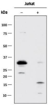
- Western blot analysis was performed on whole cell extracts (30 µg lysate) of Jurkat (Lane 1) and Jurkat treated with Staurosporine (1uM for 3 hours) (Lane 2). The blot was probed with Anti-Caspase 3 (active/pro) Monoclonal Antibody (31A1067) (Product # MA1-91637, 1µg/mL dilution) and detected by chemiluminescence using Goat anti-Mouse IgG (H+L) Superclonal™ Secondary Antibody, HRP conjugate (Product # A28177, 0.25 µg/mL, 1:4000 dilution). A 32 kDa band corresponding to Pro-Caspase 3 was observed in the cell line tested and was observed to reduce upon treatment.

- Submitted by
- Invitrogen Antibodies (provider)
- Main image

- Experimental details
- Western Blot analysis of 2mM staurosporine treated HeLa cell lysate using Active/Pro-Caspase 3 Monoclonal Antibody (31A1067) (Product # MA1-91637).

- Submitted by
- Invitrogen Antibodies (provider)
- Main image

- Experimental details
- Western Blot analysis of (A) RAW264.7 and (B) NIH-3T3 cell lysate using Active/Pro-Caspase 3 Monoclonal Antibody (31A1067) (Product # MA1-91637). Dilution: 32 µg/mL.

- Submitted by
- Invitrogen Antibodies (provider)
- Main image

- Experimental details
- Western Blot analysis of Caspase 3 (active/pro) was performed by loading Jurkat cell lysates untreated (-) or treated (+) with Etoposide. Proteins were transferred to a membrane and probed with a Caspase 3 (active/pro) Monoclonal Antibody (31A1067) (Product # MA1-91637) at a dilution of 1 µg/mL.
- Submitted by
- Invitrogen Antibodies (provider)
- Main image

- Experimental details
- Western Blot analysis of Caspase 3 (active/pro) was performed by loading HeLa cell lysate. Proteins were transferred to a membrane and probed with a Caspase 3 (active/pro) Monoclonal Antibody (31A1067) (Product # MA1-91637).

- Submitted by
- Invitrogen Antibodies (provider)
- Main image

- Experimental details
- Western Blot analysis of Caspase 3 (active/pro) was performed by loading human (A) brain, (B) heart, (C) intestine, (D) kidney, (E) liver, (F) lung, (G) muscle, (H) stomach, (I) spleen, (J) ovary, and (K) testis tissue lysates. Proteins were transferred to a membrane and probed with a Caspase 3 (active/pro) Monoclonal Antibody (31A1067) (Product # MA1-91637).

- Submitted by
- Invitrogen Antibodies (provider)
- Main image

- Experimental details
- Western Blot analysis of Caspase 3 (active/pro) was performed by loading human(Lane 1), mouse(Lane 2), and rat heart lysates(Lane 3). Proteins were transferred to a membrane and probed with a Caspase 3 (active/pro) Monoclonal Antibody (31A1067) (Product # MA1-91637).

- Submitted by
- Invitrogen Antibodies (provider)
- Main image

- Experimental details
- Western Blot analysis of Caspase 3 (active/pro) was performed by loading Jurkat cell lysates treated with and without 2 uM staurosporine. Proteins were transferred to a membrane and probed with a Caspase 3 (active/pro) Monoclonal Antibody (31A1067) (Product # MA1-91637) at a dilution of 5 µg/mL.

Supportive validation
- Submitted by
- Invitrogen Antibodies (provider)
- Main image

- Experimental details
- Immunohistochemistry (Paraffin) analysis of Caspase 3 (active/pro) in human bladder tissue using Caspase 3 (active/pro) Monoclonal Antibody (31A1067) (Product # MA1-91637) at a dilution of 1:50.

- Submitted by
- Invitrogen Antibodies (provider)
- Main image

- Experimental details
- Immunohistochemistry (Paraffin) analysis of Caspase 3 (active/pro) in human breast carcinoma tissue using Caspase 3 (active/pro) Monoclonal Antibody (31A1067) (Product # MA1-91637) at a dilution of 4 µg/mL.

- Submitted by
- Invitrogen Antibodies (provider)
- Main image

- Experimental details
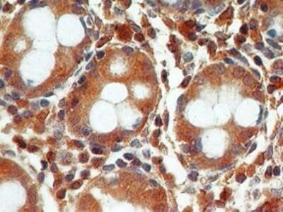
- Immunohistochemistry (Paraffin) analysis of Caspase 3 (active/pro) in human colon tissue using Caspase 3 (active/pro) Monoclonal Antibody (31A1067) (Product # MA1-91637) at a dilution of 4 µg/mL.
- Submitted by
- Invitrogen Antibodies (provider)
- Main image

- Experimental details
- Immunohistochemistry (Paraffin) analysis of Caspase 3 (active/pro) in human thymus tissue using Caspase 3 (active/pro) Monoclonal Antibody (31A1067) (Product # MA1-91637) at a dilution of 5 µg/mL. Antigen retrieval : Heat-induced antigen retrieval.

Supportive validation
- Submitted by
- Invitrogen Antibodies (provider)
- Main image

- Experimental details
- Figure 5 Viability and death of 48-h beta-arrestin 2-silenced HEK293, hGL5, and A2780 cells. Samples were transfected by increasing amount of siRNA probes targeting the beta-arrestin 2 mRNA and 48-h cell viability and death were assessed by MTT and propidium iodide. Total protein content from 3 x 10 4 70% confluent log phase cells were loaded. (A) Box and Whiskers plot representing the HEK293 cell viability under beta-arrestin 2 depletion. (B) beta-arrestin 2-silenced hGL5 cell viability. (C) beta-arrestin 2-silenced A2780 ovarian cancer cell viability. (D) Data from the beta-arrestin 2-silenced HEK293, representing cell death evaluated using propidium iodide staining. (E) hGL5 cell death evaluated by propidium iodide. (F) Cell death in beta-arrestin 2-silenced A2780 evaluated by propidium iodide. (G) Representative Western blotting images demonstrating beta-arrestin 2 depletion and pro-caspase 3 cleavage in HEK293, hGL5, and A2780 cell lysates. beta-actin was the normalizer. Brightness/contrast of Western blotting pictures were adjusted uniformly in all panels. Significantly different data distribution than mock-transfected (untransfected) cells were indicated by asterisks. Kruskal-Wallis test and Dunn's correction for multiple comparisons; * P < 0.05; ** P < 0.001; *** P < 0.0001; n = 8).

- Submitted by
- Invitrogen Antibodies (provider)
- Main image
- Experimental details
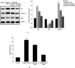
- Figure 4. Pristimerin ameliorates neuronal cell apoptosis in brain tissue homogenates of mice with sepsis-induced brain injuries. (A) Effects of pristimerin on expression levels of Bax, Bcl-2 and caspase-3, as revealed by Western blotting. (B) Effect of pristimerin on the apoptosis index, as revealed by the TUNEL assay. TUNEL: terminal deoxynucleotidyl transferase dUTP nick end-labelling. Means +- SEMs ( n = 10); ## p < 0.01 compared with positive controls; ** p < 0.01 compared with negative controls.

- Submitted by
- Invitrogen Antibodies (provider)
- Main image
- Experimental details
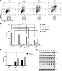
- Fig. 2 Esculetin induces apoptosis in pancreatic cancer cells: a Flow cytometric analysis of PANC-1 cells treated with 100 muM esculetin for indicated time showed temporal increase in surface expression of apoptotic marker- Annexin V indicating increased population of cells in apoptotic phase. b Percentage of PANC-1 cells exhibiting fluorescence in all four panels (healthy, early apoptosis, late apoptosis and necrosis) showing time dependent increase in apoptosis in Ecsuletin treated cells. c Percentage of cells with active APO BrdU indicating apoptosis in the absence and presence of esculetin (100 muM) as determined using TUNEL assay. d Western blot analysis showing an increase in expression of pro and active form of caspases (VC stands for vehicle control, E stands for esculetin treated sample for indicated time, CF stands for cleaved form, numerals represent time of esculetin treatment). Data represents the mean +- SD of three independent experiments. The significance was determined using ANOVA (Bonferroni's test). Key:* p < 0.05; ** p < 0.01; *** p < 0.001; **** p < 0.0001)

- Submitted by
- Invitrogen Antibodies (provider)
- Main image

- Experimental details
- Figure 2. NF-kappaB1, IL-6, MMP-13 and caspase-3 expression in the in vivo model of OA. (A) The relative protein expression levels of NF-kappaB1, IL-6, MMP-13 and caspase-3 were determined by western blot analysis in tissue samples from mice in the Sham, OA and OA + Res groups. Quantification of (B) NF-kappaB1, (C) IL-6, (D) MMP-13 and (E) caspase-3 protein expression. *P

- Submitted by
- Invitrogen Antibodies (provider)
- Main image

- Experimental details
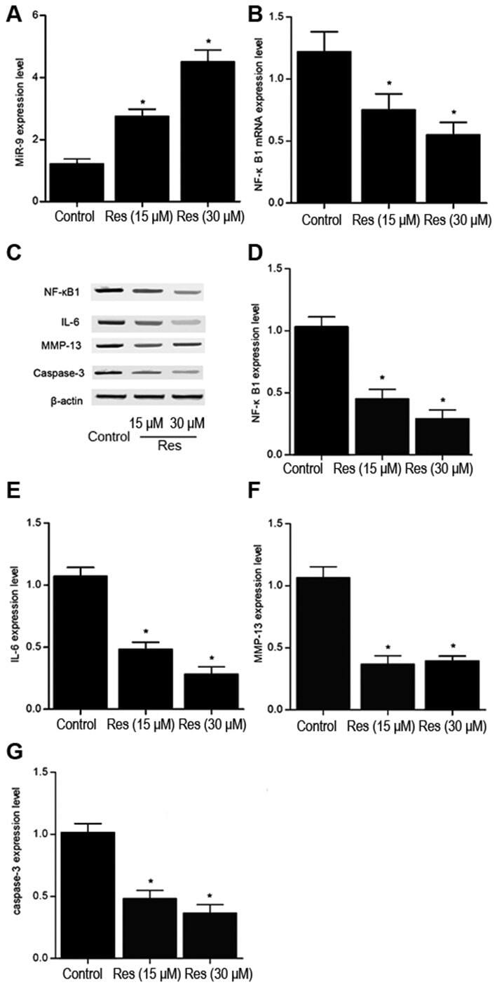
- Figure 7. Effect of Res treatment on miR-9, NF-kappaB1, IL-6, MMP-13, caspase-3 expression in mouse chondrocytes. The relative (A) miR-9 and (B) NF-kappaB1 mRNA expression levels were determined by RT-qPCR in chondrocytes following treatment with 0, 15 or 30 mM Res for 48 h. (C) The relative NF-kappaB1, IL-6, MMP-13 and caspase-3 protein expression levels were determined by western blot analysis in chondrocytes following treatment with 0, 15 or 30 mM Res for 48 h. Quantification of (D) NF-kappaB1, (E) IL-6, (F) MMP-13 and (G) caspase-3 protein expression. n=3. *P