 Explore
 Validate
 Learn
Learn Western blot
 Immunocytochemistry
Antibody data
- Antibody Data
- Antigen structure
- References [0]
- Comments [0]
- Validations
- Western blot [2]
- Immunohistochemistry [2]
Submit
Validation data
Reference
Comment
Report error
- Product number
- GTX13585 - Provider product page

- Provider
- GeneTex
- Proper citation
- GeneTex Cat#GTX13585, RRID:AB_367951
- Product name
- Caspase 3 antibody [31A1067]
- Antibody type
- Monoclonal
- Reactivity
- Human, Mouse, Rat, Hamster, Porcine
- Host
- Mouse
No comments: Submit comment
Supportive validation
- Submitted by
- GeneTex (provider)
- Main image

- Experimental details
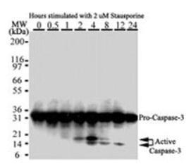
- Western blot analysis of Caspase-3 in HeLa cells. Cells were treated with 2 uM staurosporine for different time periods. Caspase-3 activation is detected in Western blots by the presence of cleavage fragments. The antibody detected both pro (full-length) and active (cleaved) protein, depending on the treatment time points. Pro Caspase-3 is detected at ~32 kDa. Active/cleaved Caspase-3 (large subunit) is detected at ~14-21 kDa as one or more bands.
- Submitted by
- GeneTex (provider)
- Main image
- Experimental details
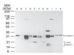
- Western blot analysis of multiple human tissues using Caspase-3 antibody at 5 μg/ml. The tissues shown are A) brain, B) heart, C) intestine, D) kidney, E) liver, F) lung, G) muscle, H) stomach, I) spleen, J) ovary, and K) testis.
Supportive validation
- Submitted by
- GeneTex (provider)
- Main image

- Experimental details
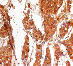
- Formalin-fixed, paraffin-embedded human breast cancer stained with Caspase-3 antibody at 4 μg/ml. Localization can be cytoplasmic and nuclear. Staining in the nucleus is considered to be an indication of active Caspase-3. In most cell types and model systems, cells with active Caspase-3 are undergoing apoptosis. Cancer/normal adjacent tissue array was used for this test.
- Submitted by
- GeneTex (provider)
- Main image
- Experimental details
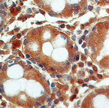
- Formalin-fixed, paraffin-embedded normal colon stained with Caspase-3 antibody at 4 μg/ml. Localization can be cytoplasmic and nuclear. Staining in the nucleus is considered to be an indication of active Caspase-3. In most cell types and model systems, cells with active Caspase-3 are undergoing apoptosis. Cancer/normal adjacent tissue array was used for this test.
