Explore
 Validate
 Learn
Learn Western blot
 Immunocytochemistry
Antibody data
- Antibody Data
- Antigen structure
- References [1]
- Comments [0]
- Validations
- Western blot [4]
- Immunohistochemistry [3]
Submit
Validation data
Reference
Comment
Report error
- Product number
- NBP2-33244 - Provider product page

- Provider
- Novus Biologicals
- Product name
- Mouse Monoclonal Caspase-3 Antibody
- Antibody type
- Monoclonal
- Description
- Protein G purified. The antibody detects both pro Caspase-3 (~32 kDa) and the large subunit of the active/cleaved form (~14-21 kDa) of Caspase-3. The large subunit of the cleaved form may appear as one or two or even as a stack of bands depending on the presence or absence of the Caspase-3 pro-domain. It is highly recommended that a maximum sensitivity ECL substrate (Femto sensitive) be used for efficient detection of this antibody in Western blot applications.
- Reactivity
- Human, Mouse, Rat, Porcine
- Host
- Mouse
- Isotype
- IgG
- Vial size
- 0.1 mg
- Storage
- Store at 4C short term. Aliquot and store at -20C long term. Avoid freeze-thaw cycles.
Submitted references Cathepsin G Inhibition by Serpinb1 and Serpinb6 Prevents Programmed Necrosis in Neutrophils and Monocytes and Reduces GSDMD-Driven Inflammation.
Burgener SS, Leborgne NGF, Snipas SJ, Salvesen GS, Bird PI, Benarafa C
Cell reports 2019 Jun 18;27(12):3646-3656.e5
Cell reports 2019 Jun 18;27(12):3646-3656.e5
No comments: Submit comment
Supportive validation
- Submitted by
- Novus Biologicals (provider)
- Main image
- Experimental details
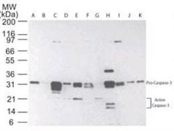
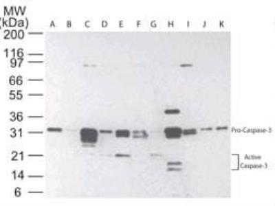
- Western Blot: Caspase-3 Antibody (31A1067) - (Pro and Active) - Azide and BSA Free [NBP2-33244] - Western blot of Caspase-3 in multiple human tissues. The tissues shown are A) brain, B) heart, C) intestine, D) kidney, E) liver, F) lung, G) muscle, H) stomach, I) spleen, J) ovary, and K) testis.
- Submitted by
- Novus Biologicals (provider)
- Main image
- Experimental details
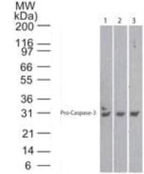
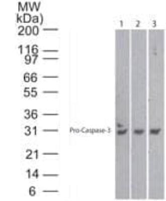
- Western Blot: Caspase-3 Antibody (31A1067) - (Pro and Active) - Azide and BSA Free [NBP2-33244] - Lanes 1, 2 and 3 demonstrate the species cross-reactivity of the antibody in human, mouse and rat heart lysate, respectively.
- Submitted by
- Novus Biologicals (provider)
- Main image

- Experimental details
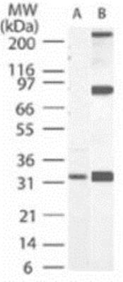
- Western Blot: Caspase-3 (Pro and Active) Antibody (31A1067) [Azide Free] [NBP2-33244] - Analysis of Caspase-3 in A) RAW and B) NIH 3T3 using 32 ug/ml.
- Submitted by
- Novus Biologicals (provider)
- Main image

- Experimental details
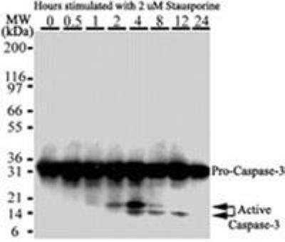
- Western Blot: Caspase-3 Antibody (31A1067) - (Pro and Active) - Azide and BSA Free [NBP2-33244] - Analysis for detection of Caspase-3 activation in HeLa cells. Cells were treated with 2mM staurosporine for different time periods. Caspase-3 activation is determined by cleavage of procaspase-3, which generates 17 and 12kDa, larger and smaller catalytic subunit, respectively.
Supportive validation
- Submitted by
- Novus Biologicals (provider)
- Main image
- Experimental details
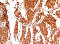
- Immunohistochemistry-Paraffin: Caspase-3 Antibody (31A1067) - (Pro and Active) - Azide and BSA Free [NBP2-33244] - Analysis in human breast cancer stained at 4ug/ml. Localization can be cytoplasmic and nuclear. Staining in the nucleus is considered to be an indication of active Caspase-3. In most cell types and model systems, cells with active Caspase-3 are undergoing apoptosis.

- Submitted by
- Novus Biologicals (provider)
- Main image
- Experimental details
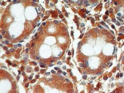
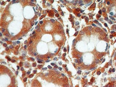
- Immunohistochemistry-Paraffin: Caspase-3 Antibody (31A1067) - (Pro and Active) - Azide and BSA Free [NBP2-33244] - Analysis in normal colon stained 4ug/ml. Localization can be cytoplasmic and nuclear. Staining in the nucleus is considered to be an indication of active Caspase-3. In most cell types and model systems, cells with active Caspase-3 are undergoing apoptosis.
- Submitted by
- Novus Biologicals (provider)
- Main image
- Experimental details
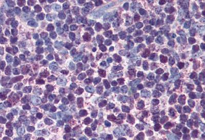
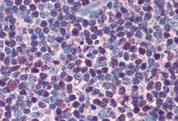
- Immunohistochemistry-Paraffin: Caspase-3 Antibody (31A1067) - (Pro and Active) - Azide and BSA Free [NBP2-33244] - Analysis in the human thymus after heat-induced antigen retrieval at 5 ug/ml.