 Explore
 Validate
 Learn
Learn Western blot
 Immunocytochemistry
Antibody data
- Antibody Data
- Antigen structure
- References [21]
- Comments [0]
- Validations
- Immunocytochemistry [4]
- Immunohistochemistry [4]
- Flow cytometry [1]
- Other assay [4]
Submit
Validation data
Reference
Comment
Report error
- Product number
- 700182 - Provider product page

- Provider
- Invitrogen Antibodies
- Product name
- Caspase 3 Recombinant Rabbit Monoclonal Antibody (9H19L2)
- Antibody type
- Monoclonal
- Antigen
- Synthetic peptide
- Description
- This antibody is predicted to react with bovine, canine, feline, hamster, mouse, primate, pufferfish, rabbit, rat, porcine and Xenopus based on sequence homology. Intact IgG appears on a non-reducing gel as ~150 kDa band and upon reduction generating a ~25 kDa light chain band and a ~50 kDa heavy chain. Recombinant rabbit monoclonal antibodies are produced using in vitro expression systems. The expression systems are developed by cloning in the specific antibody DNA sequences from immunoreactive rabbits. Then, individual clones are screened to select the best candidates for production. The advantages of using recombinant rabbit monoclonal antibodies include: better specificity and sensitivity, lot-to-lot consistency, animal origin-free formulations, and broader immunoreactivity to diverse targets due to larger rabbit immune repertoire.
- Reactivity
- Human, Mouse
- Host
- Rabbit
- Isotype
- IgG
- Antibody clone number
- 9H19L2
- Vial size
- 100 μg
- Concentration
- 0.5 mg/mL
- Storage
- Store at 4°C short term. For long term storage, store at -20°C, avoiding freeze/thaw cycles.
Submitted references HERC1 Ubiquitin Ligase Is Required for Hippocampal Learning and Memory.
Denatonium as a Bitter Taste Receptor Agonist Modifies Transcriptomic Profile and Functions of Acute Myeloid Leukemia Cells.
Silencing of soluble epoxide hydrolase 2 gene reduces H(2)O(2)-induced oxidative damage in rat intestinal epithelial IEC-6 cells via activating PI3K/Akt/GSK3β signaling pathway.
Repurposing Quinacrine for Treatment of Malignant Mesothelioma: In-Vitro Therapeutic and Mechanistic Evaluation.
Blockade of insulin receptor substrate-1 inhibits biological behavior of choroidal endothelial cells.
Parthenolide inhibits the proliferation and induces the apoptosis of human uveal melanoma cells.
Mitochondrial complex I inhibitor deguelin induces metabolic reprogramming and sensitizes vemurafenib-resistant BRAF(V600E) mutation bearing metastatic melanoma cells.
1,2,3,4,6-Penta-O-Galloyl-Beta-D-Glucopyranoside Inhibits Proliferation of Multiple Myeloma Cells Accompanied with Suppression of MYC Expression.
Effects of Lycium barbarum polysaccharides on the damage to human endometrial stromal cells induced by hydrogen peroxide.
PARP-1 depletion in combination with carbon ion exposure significantly reduces MMPs activity and overall increases TIMPs expression in cultured HeLa cells.
Expression of FADD and cFLIP(L) balances mitochondrial integrity and redox signaling to substantiate apoptotic cell death.
Identification of 1,2,3,4,6-Penta-O-galloyl-β-d-glucopyranoside as a Glycine N-Methyltransferase Enhancer by High-Throughput Screening of Natural Products Inhibits Hepatocellular Carcinoma.
Effect of memantine: A NMDA receptor blocker, on ethambutol-induced retinal injury.
Comparison of 2, 5, and 20 % O2 on the development of post-thaw human embryos.
Investigating the role of shape on the biological impact of gold nanoparticles in vitro.
Cardiac sympathetic afferent denervation attenuates cardiac remodeling and improves cardiovascular dysfunction in rats with heart failure.
Functional proteomic analysis reveals the involvement of KIAA1199 in breast cancer growth, motility and invasiveness.
Impact of diet deprivation and subsequent overallowance during gestation on mammary gland development and lactation performance.
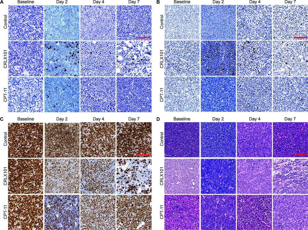
Serial diffusion MRI to monitor and model treatment response of the targeted nanotherapy CRLX101.
Detection and quantification of farnesol-induced apoptosis in difficult primary cell cultures by TaqMan protein assay.
Apoptosis-dependent acute lung injury and repair after intratracheal instillation of noradrenaline in rats.
Pérez-Villegas EM, Pérez-Rodríguez M, Negrete-Díaz JV, Ruiz R, Rosa JL, de Toledo GA, Rodríguez-Moreno A, Armengol JA
Frontiers in neuroanatomy 2020;14:592797
Frontiers in neuroanatomy 2020;14:592797
Denatonium as a Bitter Taste Receptor Agonist Modifies Transcriptomic Profile and Functions of Acute Myeloid Leukemia Cells.
Salvestrini V, Ciciarello M, Pensato V, Simonetti G, Laginestra MA, Bruno S, Pazzaglia M, De Marchi E, Forte D, Orecchioni S, Martinelli G, Bertolini F, Méndez-Ferrer S, Adinolfi E, Di Virgilio F, Cavo M, Curti A
Frontiers in oncology 2020;10:1225
Frontiers in oncology 2020;10:1225
Silencing of soluble epoxide hydrolase 2 gene reduces H(2)O(2)-induced oxidative damage in rat intestinal epithelial IEC-6 cells via activating PI3K/Akt/GSK3β signaling pathway.
Li J, Luo J, Zhang Y, Tang C, Wang J, Chen C
Cytotechnology 2020 Feb;72(1):23-36
Cytotechnology 2020 Feb;72(1):23-36
Repurposing Quinacrine for Treatment of Malignant Mesothelioma: In-Vitro Therapeutic and Mechanistic Evaluation.
Kulkarni NS, Vaidya B, Parvathaneni V, Bhanja D, Gupta V
International journal of molecular sciences 2020 Aug 31;21(17)
International journal of molecular sciences 2020 Aug 31;21(17)
Blockade of insulin receptor substrate-1 inhibits biological behavior of choroidal endothelial cells.
Qian YY, Wu HY, Liu GQ, Ren C, Lu PR, Zhang XG
International journal of ophthalmology 2019;12(9):1386-1394
International journal of ophthalmology 2019;12(9):1386-1394
Parthenolide inhibits the proliferation and induces the apoptosis of human uveal melanoma cells.
Che ST, Bie L, Li X, Qi H, Yu P, Zuo L
International journal of ophthalmology 2019;12(10):1531-1538
International journal of ophthalmology 2019;12(10):1531-1538
Mitochondrial complex I inhibitor deguelin induces metabolic reprogramming and sensitizes vemurafenib-resistant BRAF(V600E) mutation bearing metastatic melanoma cells.
Carpenter EL, Chagani S, Nelson D, Cassidy PB, Laws M, Ganguli-Indra G, Indra AK
Molecular carcinogenesis 2019 Sep;58(9):1680-1690
Molecular carcinogenesis 2019 Sep;58(9):1680-1690
1,2,3,4,6-Penta-O-Galloyl-Beta-D-Glucopyranoside Inhibits Proliferation of Multiple Myeloma Cells Accompanied with Suppression of MYC Expression.
Tseeleesuren D, Kant R, Yen CH, Hsiao HH, Chen YA
Frontiers in pharmacology 2018;9:65
Frontiers in pharmacology 2018;9:65
Effects of Lycium barbarum polysaccharides on the damage to human endometrial stromal cells induced by hydrogen peroxide.
Shan T, Shan T, Liu F, Zheng H, Li G
Molecular medicine reports 2017 Feb;15(2):879-884
Molecular medicine reports 2017 Feb;15(2):879-884
PARP-1 depletion in combination with carbon ion exposure significantly reduces MMPs activity and overall increases TIMPs expression in cultured HeLa cells.
Ghorai A, Sarma A, Chowdhury P, Ghosh U
Radiation oncology (London, England) 2016 Sep 22;11(1):126
Radiation oncology (London, England) 2016 Sep 22;11(1):126
Expression of FADD and cFLIP(L) balances mitochondrial integrity and redox signaling to substantiate apoptotic cell death.
Ranjan K, Pathak C
Molecular and cellular biochemistry 2016 Nov;422(1-2):135-150
Molecular and cellular biochemistry 2016 Nov;422(1-2):135-150
Identification of 1,2,3,4,6-Penta-O-galloyl-β-d-glucopyranoside as a Glycine N-Methyltransferase Enhancer by High-Throughput Screening of Natural Products Inhibits Hepatocellular Carcinoma.
Kant R, Yen CH, Lu CK, Lin YC, Li JH, Chen YM
International journal of molecular sciences 2016 May 4;17(5)
International journal of molecular sciences 2016 May 4;17(5)
Effect of memantine: A NMDA receptor blocker, on ethambutol-induced retinal injury.
Abdel-Hamid AA, Firgany Ael-D, Ali EM
Annals of anatomy = Anatomischer Anzeiger : official organ of the Anatomische Gesellschaft 2016 Mar;204:86-92
Annals of anatomy = Anatomischer Anzeiger : official organ of the Anatomische Gesellschaft 2016 Mar;204:86-92
Comparison of 2, 5, and 20 % O2 on the development of post-thaw human embryos.
Yang Y, Xu Y, Ding C, Khoudja RY, Lin M, Awonuga AO, Dai J, Puscheck EE, Rappolee DA, Zhou C
Journal of assisted reproduction and genetics 2016 Jul;33(7):919-27
Journal of assisted reproduction and genetics 2016 Jul;33(7):919-27
Investigating the role of shape on the biological impact of gold nanoparticles in vitro.
Tian F, Clift MJ, Casey A, Del Pino P, Pelaz B, Conde J, Byrne HJ, Rothen-Rutishauser B, Estrada G, de la Fuente JM, Stoeger T
Nanomedicine (London, England) 2015;10(17):2643-57
Nanomedicine (London, England) 2015;10(17):2643-57
Cardiac sympathetic afferent denervation attenuates cardiac remodeling and improves cardiovascular dysfunction in rats with heart failure.
Wang HJ, Wang W, Cornish KG, Rozanski GJ, Zucker IH
Hypertension (Dallas, Tex. : 1979) 2014 Oct;64(4):745-55
Hypertension (Dallas, Tex. : 1979) 2014 Oct;64(4):745-55
Functional proteomic analysis reveals the involvement of KIAA1199 in breast cancer growth, motility and invasiveness.
Jami MS, Hou J, Liu M, Varney ML, Hassan H, Dong J, Geng L, Wang J, Yu F, Huang X, Peng H, Fu K, Li Y, Singh RK, Ding SJ
BMC cancer 2014 Mar 15;14:194
BMC cancer 2014 Mar 15;14:194
Impact of diet deprivation and subsequent overallowance during gestation on mammary gland development and lactation performance.
Farmer C, Palin MF, Martel-Kennes Y
Journal of animal science 2014 Jan;92(1):141-51
Journal of animal science 2014 Jan;92(1):141-51
Serial diffusion MRI to monitor and model treatment response of the targeted nanotherapy CRLX101.
Ng TS, Wert D, Sohi H, Procissi D, Colcher D, Raubitschek AA, Jacobs RE
Clinical cancer research : an official journal of the American Association for Cancer Research 2013 May 1;19(9):2518-27
Clinical cancer research : an official journal of the American Association for Cancer Research 2013 May 1;19(9):2518-27
Detection and quantification of farnesol-induced apoptosis in difficult primary cell cultures by TaqMan protein assay.
Pfister C, Pfrommer H, Tatagiba MS, Roser F
Apoptosis : an international journal on programmed cell death 2013 Apr;18(4):452-66
Apoptosis : an international journal on programmed cell death 2013 Apr;18(4):452-66
Apoptosis-dependent acute lung injury and repair after intratracheal instillation of noradrenaline in rats.
Uhal BD, Rayford H, Zhuang J, Li X, Laukka J, Soledad-Conrad V
Experimental physiology 2003 Mar;88(2):269-75
Experimental physiology 2003 Mar;88(2):269-75
No comments: Submit comment
Supportive validation
- Submitted by
- Invitrogen Antibodies (provider)
- Main image
- Experimental details
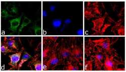
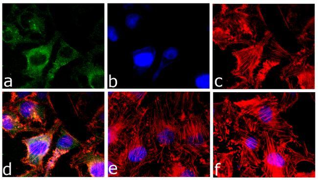
- Immunofluorescence analysis of Caspase-3 was done on 70% confluent log phase HeLa cells treated with 5 uM of Staurosporine for 16 hours. The cells were fixed with 4% paraformaldehyde for 10 minutes, permeabilized with 0.1% Triton™ X-100 for 10 minutes, and blocked with 5% BSA for 1 hour at room temperature. The cells were labeled with Caspase-3 (9H19L2) Recombinant Rabbit Monoclonal Antibody (Product # 700182) at 1 µg/mL in 0.1% BSA and incubated for 3 hours at room temperature and then labeled with Goat anti-Rabbit IgG (H+L) Superclonal™ Secondary Antibody, Alexa Fluor® 488 conjugate (Product # A27034) at a dilution of 1:2000 for 45 minutes at room temperature (Panel a: green). Nuclei (Panel b: blue) were stained with SlowFade® Gold Antifade Mountant with DAPI (Product # S36938). F-actin (Panel c: red) was stained with Alexa Fluor® 555 Rhodamine Phalloidin (Product # R415, 1:300). Panel d is a merged image showing Cytoplasmic localization. Panel e is untreated cell showing no signal. Panel f is a no primary antibody control. The images were captured at 60X magnification

- Submitted by
- Invitrogen Antibodies (provider)
- Main image
- Experimental details
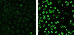
- Immunofluorescent analysis of Caspase-3 in A549 cells treated with staurosporine (right) and untreated (left) using a Caspase-3 recombinant rabbit monoclonal antibody (Product # 700182) at a dilution of 5 µg/mL followed by detection using an Alexa Fluor 488-conjugated goat anti-rabbit secondary antibody at a dilution of 1:1000.

- Submitted by
- Invitrogen Antibodies (provider)
- Main image
- Experimental details
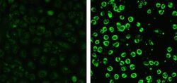
- Immunofluorescent analysis of Caspase-3 in A549 cells treated with staurosporine (right) and untreated (left) using a Caspase-3 recombinant rabbit monoclonal antibody (Product # 700182) at a dilution of 5 µg/mL followed by detection using an Alexa Fluor 488-conjugated goat anti-rabbit secondary antibody at a dilution of 1:1000.
- Submitted by
- Invitrogen Antibodies (provider)
- Main image

- Experimental details
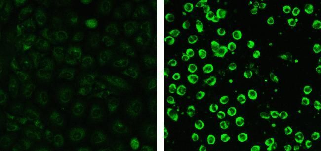
- Immunofluorescence analysis of Caspase-3 was done on 70% confluent log phase HeLa cells treated with 5 uM of Staurosporine for 16 hours. The cells were fixed with 4% paraformaldehyde for 10 minutes, permeabilized with 0.1% Triton™ X-100 for 10 minutes, and blocked with 5% BSA for 1 hour at room temperature. The cells were labeled with Caspase-3 (9H19L2) Recombinant Rabbit Monoclonal Antibody (Product # 700182) at 1 µg/mL in 0.1% BSA and incubated for 3 hours at room temperature and then labeled with Goat anti-Rabbit IgG (Heavy Chain) Superclonal™ Secondary Antibody, Alexa Fluor® 488 conjugate (Product # A27034) at a dilution of 1:2000 for 45 minutes at room temperature (Panel a: green). Nuclei (Panel b: blue) were stained with SlowFade® Gold Antifade Mountant with DAPI (Product # S36938). F-actin (Panel c: red) was stained with Alexa Fluor® 555 Rhodamine Phalloidin (Product # R415, 1:300). Panel d is a merged image showing Cytoplasmic localization. Panel e is untreated cell showing no signal. Panel f is a no primary antibody control. The images were captured at 60X magnification
Supportive validation
- Submitted by
- Invitrogen Antibodies (provider)
- Main image

- Experimental details
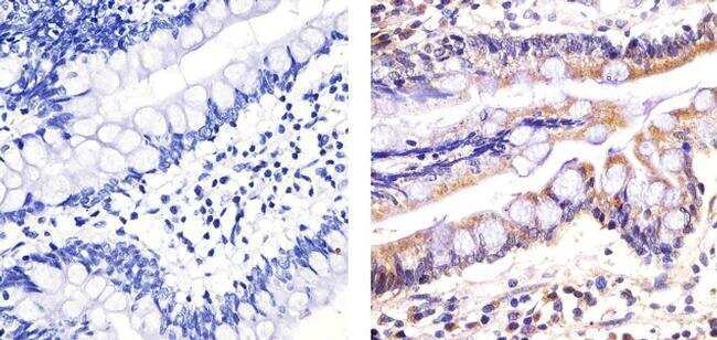
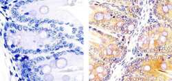
- Immunohistochemistry analysis of CASP3 showing staining in the cytoplasm of paraffin-embedded human colon tissue (right) compared to a negative control without primary antibody (left). To expose target proteins, antigen retrieval was performed using 10mM sodium citrate (pH 6.0), microwaved for 8-15 min. Following antigen retrieval, tissues were blocked in 3% H2O2-methanol for 15 min at room temperature, washed with ddH2O and PBS, and then probed with a CASP3 (9H19L2) Recombinant Rabbit Monoclonal Antibody (Product # 700182) diluted in 3% BSA-PBS at a dilution of 1:20 overnight at 4°C in a humidified chamber. Tissues were washed extensively in PBST and detection was performed using an HRP-conjugated secondary antibody followed by colorimetric detection using a DAB kit. Tissues were counterstained with hematoxylin and dehydrated with ethanol and xylene to prep for mounting.
- Submitted by
- Invitrogen Antibodies (provider)
- Main image
- Experimental details
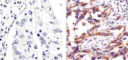
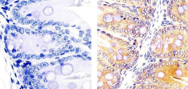
- Immunohistochemistry analysis of CASP3 showing staining in the cytoplasm of paraffin-embedded human lung adenocarcinoma tissue (right) compared to a negative control without primary antibody (left). To expose target proteins, antigen retrieval was performed using 10mM sodium citrate (pH 6.0), microwaved for 8-15 min. Following antigen retrieval, tissues were blocked in 3% H2O2-methanol for 15 min at room temperature, washed with ddH2O and PBS, and then probed with a CASP3 (9H19L2) Recombinant Rabbit Monoclonal Antibody (Product # 700182) diluted in 3% BSA-PBS at a dilution of 1:50 overnight at 4ºC in a humidified chamber. Tissues were washed extensively in PBST and detection was performed using an HRP-conjugated secondary antibody followed by colorimetric detection using a DAB kit. Tissues were counterstained with hematoxylin and dehydrated with ethanol and xylene to prep for mounting.
- Submitted by
- Invitrogen Antibodies (provider)
- Main image
- Experimental details
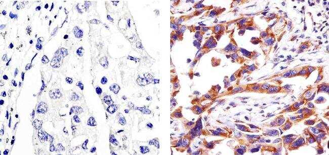
- Immunohistochemistry analysis of CASP3 showing staining in the cytoplasm of paraffin-embedded mouse colon tissue (right) compared to a negative control without primary antibody (left). To expose target proteins, antigen retrieval was performed using 10mM sodium citrate (pH 6.0), microwaved for 8-15 min. Following antigen retrieval, tissues were blocked in 3% H2O2-methanol for 15 min at room temperature, washed with ddH2O and PBS, and then probed with a CASP3 (9H19L2) Recombinant Rabbit Monoclonal Antibody (Product # 700182) diluted in 3% BSA-PBS at a dilution of 1:20 overnight at 4ºC in a humidified chamber. Tissues were washed extensively in PBST and detection was performed using an HRP-conjugated secondary antibody followed by colorimetric detection using a DAB kit. Tissues were counterstained with hematoxylin and dehydrated with ethanol and xylene to prep for mounting.
- Submitted by
- Invitrogen Antibodies (provider)
- Main image
- Experimental details
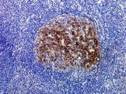
- Immunohistochemistry analysis of Caspase-3 in formalin-fixed, paraffin-embedded human tonsil tissue using a Caspase-3 monoclonal antibody (Product # 700182) at a dilution of 1 µg/mL. Staining was visualized using DAB and images were taken at a magnification of 20x. Results show cytoplasmic staining of proliferating cells in the germinal center area.

Supportive validation
- Submitted by
- Invitrogen Antibodies (provider)
- Main image
- Experimental details
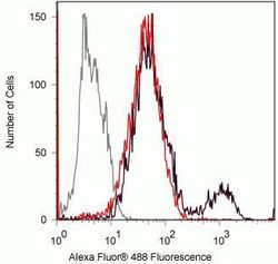
- Flow cytometry analysis of Caspase-3 in Jurkat cells incubated with 10uM camptothecin for 4hr (black) or untreated (red) using a Caspase-3 recombinant rabbit monoclonal antibody (Product # 700182) at a dilution of 0.5ug. Cells were fixed and permeabilized using FIX & PERM (Product # GAS004) reagent, and detection was performed using an Alexa Fluor 488 goat anti-rabbit IgG compared to a control without primary antibody (gray).

Supportive validation
- Submitted by
- Invitrogen Antibodies (provider)
- Main image

- Experimental details
- NULL
- Submitted by
- Invitrogen Antibodies (provider)
- Main image
- Experimental details
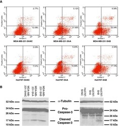
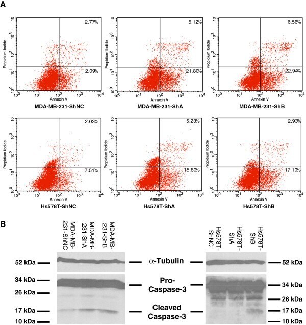
- Figure 3 KIAA1199 Knockdown enhanced apoptosis in vitro . A) Flow cytometry analysis shows a large increase in the percentage of cells programmed for apoptosis in MDA-MB-231-ShA, MDA-MB-231-ShB, Hs578T-ShA and Hs578T-ShB cells comparing to the corresponding negative controls. B) Confirmation of the results of Flow cytometry analysis by Western blot (single experiment). Caspase-3 activation is detected in Western blots by the presence of cleavage fragments. The antibody detects both pro (full-length) and active (cleaved) protein. The increased representation of cleaved caspase-3 in KIAA1199 knockdown cells compared to the control cells is qualitatively shown in MDA-MB-231 (left panel) and Hs578T (right panel) cells.
- Submitted by
- Invitrogen Antibodies (provider)
- Main image
- Experimental details
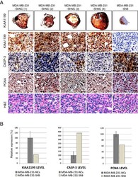
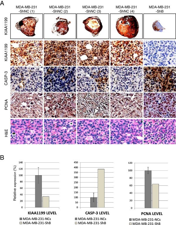
- Figure 5 Immunohistochemical studies. A) Very low KIAA1199 immunostaining (first row) in MDA-MB-231-ShB tumor comparing to the controls (x4). Representative illustration of immunohistochemical studies (x100 magnifications) shows the higher expression level of KIAA1199 (brown staining cells in the second row), lower apoptosis activity (CASP3, third row) and higher proliferation activity (PCNA, fourth row) than the MDA-MB-231-ShB tumor. B) Evaluation of the expression of protein markers by calculation of immunostaining index using the Metamorph software; graphs from left to right show the relative expression of KIAA1199, CASP3 and PCNA in control versus KIAA1199 knockdown tumor sections.
- Submitted by
- Invitrogen Antibodies (provider)
- Main image
- Experimental details
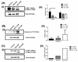
- Figure 8 ( A )-i Western blots representing the expression level of two subtypes (1 and 2) of LC3B protein post QA treatment (5- and 10 uM, 24 h) in MSTO-211H cells. It can be clearly seen that LC3B-1 is downregulated and LC3B-2 is upregulated in MSTO-211H cells, indicating successful inhibition of autophagy. ( A )-ii Plot represents band intensity ratio for protein and B-actin for LC3B protein subtypes in MSTO-211H cells. A significant difference can be seen for LC3B-1 downregulation for QA treated cells as compared to control cells. ( B )-i Western blot representing expression level of caspase-3, which is overexpressed in apoptotic events. It can be clearly seen that QA treatment (2.5- and 5 uM, 72 h) induces levels of caspase-3, indicating successful apoptosis. ( B )-ii Plot represents band intensity ratio for protein and B-actin for caspase-3 in MSTO-211H cells. An upregulation of Caspase-3 marker can be seen as QA concentration is increased, indicating induction of apoptosis. ( C )-i Blots represent LC3B expression levels in H2452 cells for control cells and QA treated cells. A clear induction in LC3B-2 levels are observed whereas LC3B-1 levels are seen to be downregulated. ( C )-ii Plot represents band intensity ratio for protein and B-actin for LC3B-2 in MSTO-211H cells. Data represents mean +- SD ( n = 3), * p < 0.05, compared between treatment and control groups, as indicated.
