Explore
 Validate
 Learn
Learn Western blot
 Immunocytochemistry
Antibody data
- Antibody Data
- Antigen structure
- References [1]
- Comments [0]
- Validations
- Western blot [1]
- Immunohistochemistry [5]
Submit
Validation data
Reference
Comment
Report error
- Product number
- NBP1-90125 - Provider product page

- Provider
- Novus Biologicals
- Proper citation
- Novus Cat#NBP1-90125, RRID:AB_11040670
- Product name
- Rabbit Polyclonal Caspase-3 Antibody
- Antibody type
- Polyclonal
- Description
- Immunogen affinity purified. Specificity of human Caspase-3 antibody verified on a Protein Array containing target protein plus 383 other non-specific proteins.
- Reactivity
- Human, Rat
- Host
- Rabbit
- Isotype
- IgG
- Vial size
- 0.1 ml
- Storage
- Store at 4C short term. Aliquot and store at -20C long term. Avoid freeze-thaw cycles.
Submitted references Photoreceptor damage induced by low-intensity light: model of retinal degeneration in mammals.
Contín MA, Arietti MM, Benedetto MM, Bussi C, Guido ME
Molecular vision 2013;19:1614-25
Molecular vision 2013;19:1614-25
No comments: Submit comment
Supportive validation
- Submitted by
- Novus Biologicals (provider)
- Main image

- Experimental details
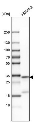
- Western Blot: Caspase-3 Antibody [NBP1-90125] - Analysis in human cell line HDLM-2.
Supportive validation
- Submitted by
- Novus Biologicals (provider)
- Main image
- Experimental details
- Immunohistochemistry-Paraffin: Caspase-3 Antibody [NBP1-90125] - HUVEC cells were fixed for 10 minutes using 10% formalin and then permeabilized for 5 minutes using 1X TBS + 0.5% Triton X-100. The cells were incubated with CD31/PECAM-1 Antibody at 5 ug/mL overnight at 4C and detected with an anti-rabbit Dylight 488 (Green) at a 1:500 dilution. Alpha tubulin (DM1A) NB100-690 was used as a co-stain at a 1:1000 dilution and detected with an anti-mouse Dylight 550 (Red) at a 1:500 dilution. Nuclei were counterstained with DAPI (Blue). Cells were imaged using a 40X objective.
- Submitted by
- Novus Biologicals (provider)
- Main image
- Experimental details
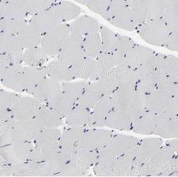
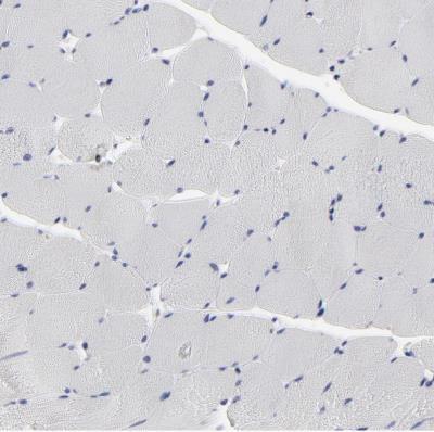
- Immunohistochemistry-Paraffin: Caspase-3 Antibody [NBP1-90125] - Staining of human skeletal muscle shows no cytoplasmic positivity in myocytes as expected.
- Submitted by
- Novus Biologicals (provider)
- Main image

- Experimental details
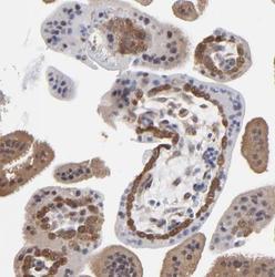
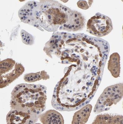
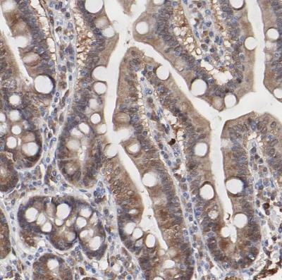
- Immunohistochemistry-Paraffin: Caspase-3 Antibody [NBP1-90125] - Staining of human appendix shows moderate to strong cytoplasmic positivity in glandular cells.
- Submitted by
- Novus Biologicals (provider)
- Main image
- Experimental details
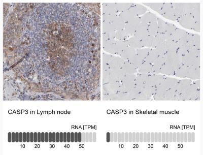
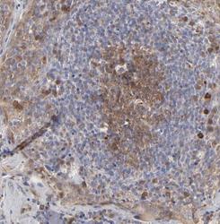
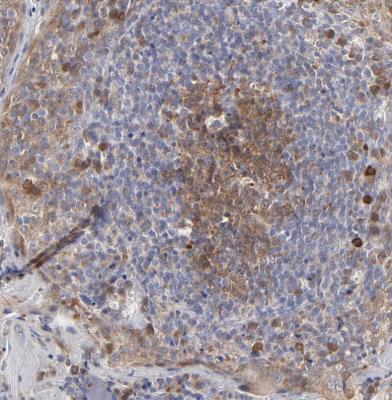
- Immunohistochemistry-Paraffin: Caspase-3 Antibody [NBP1-90125] - Staining of human lymph node shows moderate to strong cytoplasmic positivity in germinal center cells.
- Submitted by
- Novus Biologicals (provider)
- Main image

- Experimental details
- Immunohistochemistry-Paraffin: Caspase-3 Antibody [NBP1-90125] - Analysis in human lymph node and skeletal muscle tissues. Corresponding CASP3 RNA-seq data are presented for the same tissues.