Explore
 Validate
 Learn
Learn Western blot
Antibody data
- Antibody Data
- Antigen structure
- References [9]
- Comments [0]
- Validations
- Western blot [4]
- Immunocytochemistry [4]
- Immunoprecipitation [1]
- Immunohistochemistry [2]
- Flow cytometry [1]
- Other assay [3]
Submit
Validation data
Reference
Comment
Report error
- Product number
- MA5-11521 - Provider product page

- Provider
- Invitrogen Antibodies
- Product name
- Caspase 3 Monoclonal Antibody (3CSP03 (4.1.18))
- Antibody type
- Monoclonal
- Antigen
- Recombinant full-length protein
- Description
- MA5-11521 targets Caspase 3 in immunohistochemistry (paraffin), immunoprecipitation, and Western blot applications and shows reactivity with Human samples. The MA5-11521 immunogen is recombinant full length human Caspase 3 protein.
- Reactivity
- Human
- Host
- Mouse
- Isotype
- IgG
- Antibody clone number
- 3CSP03 (4.1.18)
- Vial size
- 500 μL
- Concentration
- 0.2 mg/mL
- Storage
- 4°C
Submitted references MYBL2 in synergy with CDC20 promotes the proliferation and inhibits apoptosis of gastric cancer cells.
Pulsed focused ultrasound enhances the therapeutic effect of mesenchymal stromal cell-derived extracellular vesicles in acute kidney injury.
Effects of caffeine on neuronal apoptosis in neonatal hypoxic-ischemic brain injury.
Combination of molecular alterations and smoking intensity predicts bladder cancer outcome: a report from the Los Angeles Cancer Surveillance Program.
Human papillomavirus in oral atrophic lichen planus lesions.
Increased vein wall apoptosis in varicose vein disease is related to venous hypertension.
Immunohistochemical analysis of non-small cell lung cancer: correlation with clinical parameters and prognosis.
Caspase-3 and caspase-6 in ductal breast carcinoma: a descriptive study.
Caspase-3 and caspase-6 in ductal breast carcinoma: a descriptive study.
Deng Q, Wu L, Li Y, Zou L
Advances in clinical and experimental medicine : official organ Wroclaw Medical University 2021 Sep;30(9):957-966
Advances in clinical and experimental medicine : official organ Wroclaw Medical University 2021 Sep;30(9):957-966
Pulsed focused ultrasound enhances the therapeutic effect of mesenchymal stromal cell-derived extracellular vesicles in acute kidney injury.
Ullah M, Liu DD, Rai S, Razavi M, Concepcion W, Thakor AS
Stem cell research & therapy 2020 Sep 14;11(1):398
Stem cell research & therapy 2020 Sep 14;11(1):398
Effects of caffeine on neuronal apoptosis in neonatal hypoxic-ischemic brain injury.
Kilicdag H, Daglioglu YK, Erdogan S, Zorludemir S
The journal of maternal-fetal & neonatal medicine : the official journal of the European Association of Perinatal Medicine, the Federation of Asia and Oceania Perinatal Societies, the International Society of Perinatal Obstetricians 2014 Sep;27(14):1470-5
The journal of maternal-fetal & neonatal medicine : the official journal of the European Association of Perinatal Medicine, the Federation of Asia and Oceania Perinatal Societies, the International Society of Perinatal Obstetricians 2014 Sep;27(14):1470-5
Combination of molecular alterations and smoking intensity predicts bladder cancer outcome: a report from the Los Angeles Cancer Surveillance Program.
Mitra AP, Castelao JE, Hawes D, Tsao-Wei DD, Jiang X, Shi SR, Datar RH, Skinner EC, Stein JP, Groshen S, Yu MC, Ross RK, Skinner DG, Cortessis VK, Cote RJ
Cancer 2013 Feb 15;119(4):756-65
Cancer 2013 Feb 15;119(4):756-65
Human papillomavirus in oral atrophic lichen planus lesions.
Mattila R, Rautava J, Syrjänen S
Oral oncology 2012 Oct;48(10):980-984
Oral oncology 2012 Oct;48(10):980-984
Increased vein wall apoptosis in varicose vein disease is related to venous hypertension.
Filis K, Kavantzas N, Isopoulos T, Antonakis P, Sigalas P, Vavouranakis E, Sigala F
European journal of vascular and endovascular surgery : the official journal of the European Society for Vascular Surgery 2011 Apr;41(4):533-9
European journal of vascular and endovascular surgery : the official journal of the European Society for Vascular Surgery 2011 Apr;41(4):533-9
Immunohistochemical analysis of non-small cell lung cancer: correlation with clinical parameters and prognosis.
Yoo J, Jung JH, Lee MA, Seo KJ, Shim BY, Kim SH, Cho DG, Ahn MI, Kim CH, Cho KD, Kang SJ, Kim HK
Journal of Korean medical science 2007 Apr;22(2):318-25
Journal of Korean medical science 2007 Apr;22(2):318-25
Caspase-3 and caspase-6 in ductal breast carcinoma: a descriptive study.
Blázquez S, Sirvent JJ, Olona M, Aguilar C, Pelegri A, Garcia JF, Palacios J
Histology and histopathology 2006 Dec;21(12):1321-9
Histology and histopathology 2006 Dec;21(12):1321-9
Caspase-3 and caspase-6 in ductal breast carcinoma: a descriptive study.
Blázquez S, Sirvent JJ, Olona M, Aguilar C, Pelegri A, Garcia JF, Palacios J
Histology and histopathology 2006 Dec;21(12):1321-9
Histology and histopathology 2006 Dec;21(12):1321-9
No comments: Submit comment
Supportive validation
- Submitted by
- Invitrogen Antibodies (provider)
- Main image
- Experimental details
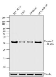
- Western blot analysis was performed on whole cell extracts (30 µg lysate) of HEL 92.1.7 (Lane 1), K562 (Lane 2), NTERA-2 (Lane 3), and MDA-MB-231 (Lane 4). The blots were probed with Anti-Caspase-3 Mouse Monoclonal Antibody (Product # MA5-11521, 1-2 µg/mL) and detected by chemiluminescence using Goat anti-Mouse IgG (H+L) Secondary Antibody, HRP conjugate (Product # 62-6520, 1:4000 dilution). A 31 kDa band corresponding to Caspase-3 was observed across cell lines tested. Known quantity of protein samples were electrophoresed using Novex® NuPAGE® 12 % Bis-Tris gel (Product # NP0342BOX), XCell SureLock™ Electrophoresis System (Product # EI0002) and Novex® Sharp Pre-Stained Protein Standard (Product # LC5800). Resolved proteins were then transferred onto a nitrocellulose membrane with iBlot® 2 Dry Blotting System (Product # IB21001). The membrane was probed with the relevant primary and secondary Antibody following blocking with 5 % skimmed milk. Chemiluminescent detection was performed using Pierce™ ECL Western Blotting Substrate (Product # 32106).
- Submitted by
- Invitrogen Antibodies (provider)
- Main image
- Experimental details
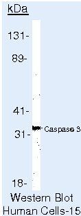
- Western blot of Caspase 3 using Caspase 3 Monoclonal Antibody (Product # MA5-11521) on Jurkat Cells.
- Submitted by
- Invitrogen Antibodies (provider)
- Main image
- Experimental details
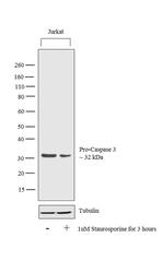
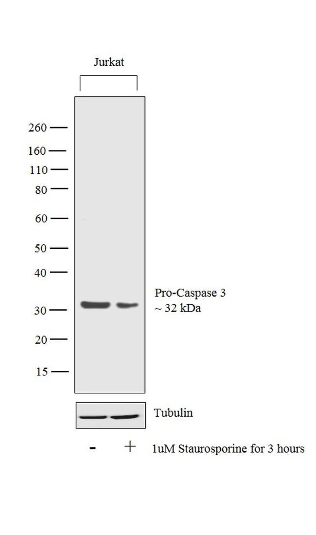
- Western blot analysis was performed on whole cell extracts (30 µg lysate) of Jurkat (Lane 1) and Jurkat treated with Staurosporine (1uM for 3 hours) (Lane 2). The blot was probed with Anti-Caspase 3 Monoclonal Antibody (3CSP03(4.1.18)) (Product # MA5-11521, 1µg/mL dilution) and detected by chemiluminescence using Goat anti-Mouse IgG (H+L) Superclonal™ Secondary Antibody, HRP conjugate (Product # A28177, 0.25 µg/mL, 1:4000 dilution). A 32 kDa band corresponding to Caspase 3 was observed in the cell line tested and was observed to reduce upon treatment.
- Submitted by
- Invitrogen Antibodies (provider)
- Main image

- Experimental details
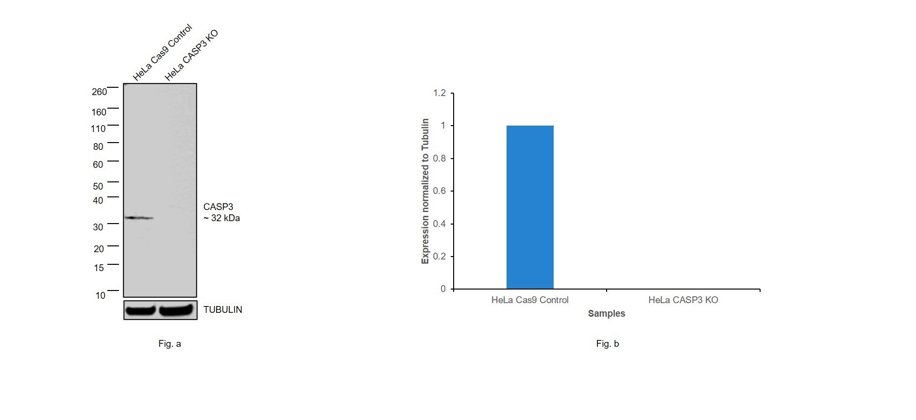
- Knockout of CASP3 was achieved by CRISPR-Cas9 genome editing using LentiArray™ Lentiviral sgRNA (Product # A32042, Assay ID CRISPR797315_LV) and LentiArray Cas9 Lentivirus (Product # A32064). Western blot analysis of CASP3 was performed by loading 30 µg of HeLa Cas9 (Lane 1) andHeLa CASP3 KO (Lane 2) whole cell extracts. The samples were electrophoresed using NuPAGE™ Novex™ 4-12% Bis-Tris Protein Gel (Product # NP0322BOX). Resolved proteins were then transferred onto a nitrocellulose membrane (Product # IB23001) by iBlot® 2 Dry Blotting System (Product # IB21001). The blot was probed with Anti-Caspase 3 Monoclonal Antibody (3CSP03 (4.1.18)) (Product # MA5-11521, 1 µg/mL dilution) and Goat anti-Mouse IgG (H+L) Superclonal™ Recombinant Secondary Antibody, HRP (Product # A28177, 1:5,000 dilution) using the iBright FL 1000 (Product # A32752). Chemiluminescent detection was performed using SuperSignal™ West Dura Extended Duration Substrate (Product # 34076). Loss of signal upon CRISPR mediated knockout (KO) using the LentiArray™ CRISPR product line confirms that antibody is specific to CASP3.
Supportive validation
- Submitted by
- Invitrogen Antibodies (provider)
- Main image

- Experimental details
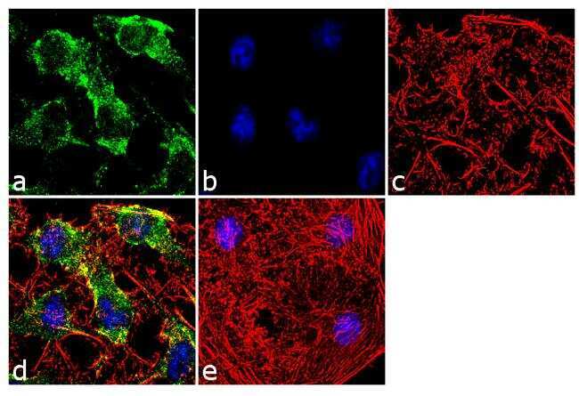
- Immunofluorescence analysis of Caspase 3 was done on 70% confluent log phase HeLa cells. The cells were fixed with 4% paraformaldehyde for 10 minutes, permeabilized with 0.1% Triton™ X-100 for 10 minutes, and blocked with 1% BSA for 1 hour at room temperature. The cells were labeled with Caspase 3 (3CSP03 (4.1.18)) Mouse Monoclonal Antibody (Product # MA5-11521) at 2 µg/mL in 0.1% BSA and incubated for 3 hours at room temperature and then labeled with Goat anti-Mouse IgG (H+L) Superclonal™ Secondary Antibody, Alexa Fluor® 488 conjugate (Product # A28175) at a dilution of 1:2000 for 45 minutes at room temperature (Panel a: green). Nuclei (Panel b: blue) were stained with SlowFade® Gold Antifade Mountant with DAPI (Product # S36938). F-actin (Panel c: red) was stained with Alexa Fluor® 555 Rhodamine Phalloidin (Product # R415, 1:300).Panel d is a merged image showing cytoplasmic localization. Panel e is a no primary antibody control. The images were captured at 60X magnification.
- Submitted by
- Invitrogen Antibodies (provider)
- Main image
- Experimental details
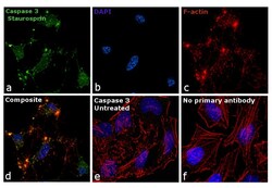
- Immunofluorescence analysis of Caspase 3 was performed using 70% confluent log phase HeLa cells treated with 1 µg/mL Staurosporine for 5 hours. The cells were fixed with 4% paraformaldehyde for 10 minutes, permeabilized with 0.1% Triton™ X-100 for 10 minutes, and blocked with 1% BSA for 1 hour at room temperature. The cells were labeled with Caspase 3 Monoclonal Antibody (3CSP03 (4.1.18)) (product # MA5-24681) at 2µg/mL in 0.1% BSA, incubated overnight at 4 degree Celsius and then labeled with Goat anti-Mouse IgG (H+L) Superclonal™ Secondary Antibody, Alexa Fluor® 488 conjugate (Product # A28175) at a dilution of 1:2000 for 45 minutes at room temperature (Panel a: green). Nuclei (Panel b: blue) were stained with SlowFade® Gold Antifade Mountant with DAPI (Product # S36938). F-actin (Panel c: red) was stained with Rhodamine Phalloidin (Product # R415, 1:300). Panel d represents the merged image showing cytoplasmic localization upon Staurosporine treatment. Panel e shows untreated cells without any staining. Panel f represents control cells with no primary antibody to assess background. The images were captured at 60X magnification.

- Submitted by
- Invitrogen Antibodies (provider)
- Main image

- Experimental details
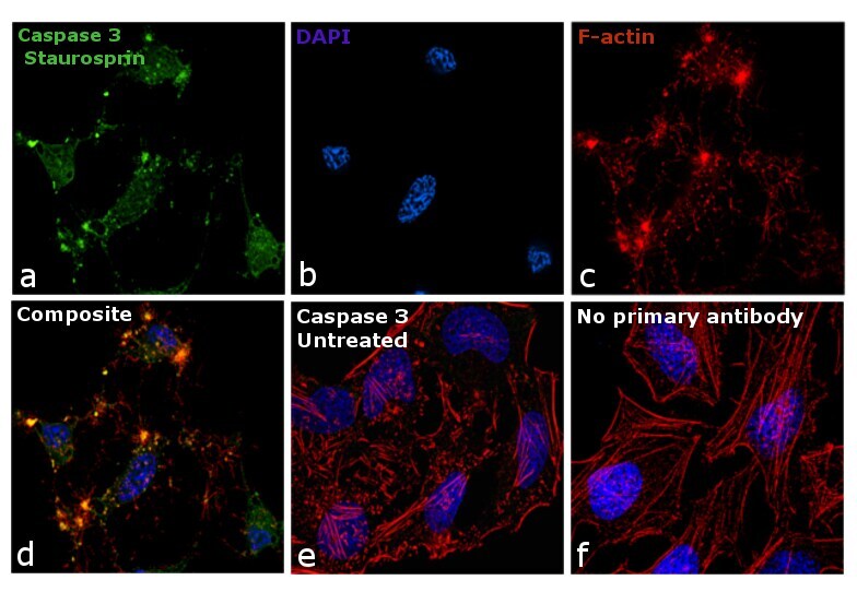
- Immunofluorescence analysis of Caspase 3 was performed using 70% confluent log phase HeLa cells treated with 1 µg/mL Staurosporine for 5 hours. The cells were fixed with 4% paraformaldehyde for 10 minutes, permeabilized with 0.1% Triton™ X-100 for 10 minutes, and blocked with 1% BSA for 1 hour at room temperature. The cells were labeled with Caspase 3 Monoclonal Antibody (3CSP03 (4.1.18)) (product # MA5-24681) at 2µg/mL in 0.1% BSA, incubated overnight at 4 degree Celsius and then labeled with Goat anti-Mouse IgG (H+L) Superclonal™ Secondary Antibody, Alexa Fluor® 488 conjugate (Product # A28175) at a dilution of 1:2000 for 45 minutes at room temperature (Panel a: green). Nuclei (Panel b: blue) were stained with SlowFade® Gold Antifade Mountant with DAPI (Product # S36938). F-actin (Panel c: red) was stained with Rhodamine Phalloidin (Product # R415, 1:300). Panel d represents the merged image showing cytoplasmic localization upon Staurosporine treatment. Panel e shows untreated cells without any staining. Panel f represents control cells with no primary antibody to assess background. The images were captured at 60X magnification.
- Submitted by
- Invitrogen Antibodies (provider)
- Main image

- Experimental details
- Immunofluorescence analysis of Caspase 3 was done on 70% confluent log phase HeLa cells. The cells were fixed with 4% paraformaldehyde for 10 minutes, permeabilized with 0.1% Triton™ X-100 for 10 minutes, and blocked with 1% BSA for 1 hour at room temperature. The cells were labeled with Caspase 3 (3CSP03 (4.1.18)) Mouse Monoclonal Antibody (Product # MA5-11521) at 2 µg/mL in 0.1% BSA and incubated for 3 hours at room temperature and then labeled with Goat anti-Mouse IgG (H+L) Superclonal™ Secondary Antibody, Alexa Fluor® 488 conjugate (Product # A28175) at a dilution of 1:2000 for 45 minutes at room temperature (Panel a: green). Nuclei (Panel b: blue) were stained with SlowFade® Gold Antifade Mountant with DAPI (Product # S36938). F-actin (Panel c: red) was stained with Alexa Fluor® 555 Rhodamine Phalloidin (Product # R415, 1:300).Panel d is a merged image showing cytoplasmic localization. Panel e is a no primary antibody control. The images were captured at 60X magnification.

Supportive validation
- Submitted by
- Invitrogen Antibodies (provider)
- Main image

- Experimental details
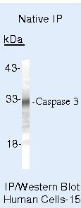
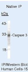
- Immunoprecipitation of Caspase 3 using Caspase 3 Monoclonal Antibody (Product # MA5-11521) on Native Human Jurkat Cells.
Supportive validation
- Submitted by
- Invitrogen Antibodies (provider)
- Main image
- Experimental details
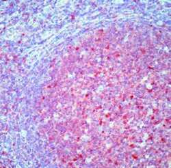
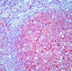
- Formalin-fixed, paraffin-embedded human tonsil stained with Caspase 3 antibody using peroxidase-conjugate and AEC chromogen. Note cytoplasmic and nuclear staining of lymphoid cells.
- Submitted by
- Invitrogen Antibodies (provider)
- Main image
- Experimental details
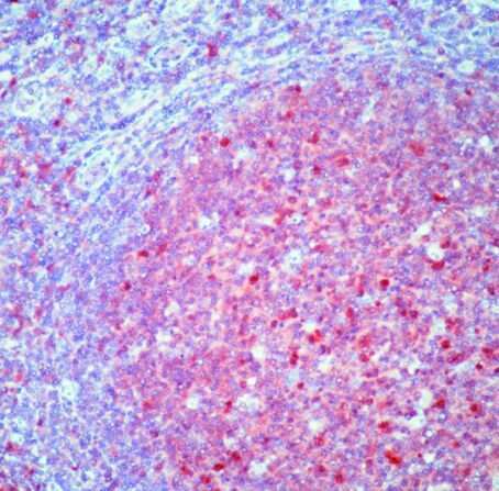
- Formalin-fixed, paraffin-embedded human tonsil stained with Caspase 3 antibody using peroxidase-conjugate and AEC chromogen. Note cytoplasmic and nuclear staining of lymphoid cells.

Supportive validation
- Submitted by
- Invitrogen Antibodies (provider)
- Main image
- Experimental details
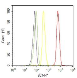
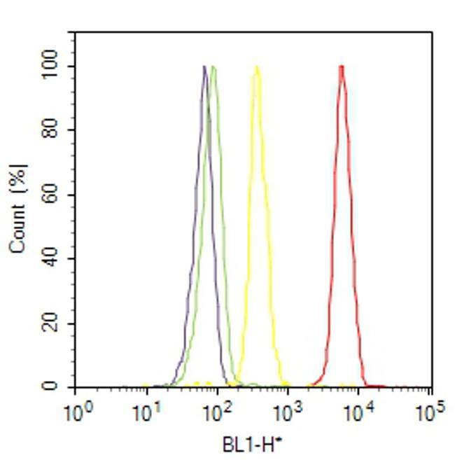
- Flow cytometry analysis of Caspase 3 was done on HEL cells. Cells were fixed with 70% ethanol for 10 minutes, permeabilized with 0.25% Triton™ X-100 for 20 minutes, and blocked with 5% BSA for 30 minutes at room temperature. Cells were labeled with Caspase 3 Mouse Monoclonal Antibody (MA511521, red histogram) or with mouse isotype control (yellow histogram) at 3-5 ug/million cells in 2.5% BSA. After incubation at room temperature for 2 hours, the cells were labeled with Alexa Fluor® 488 Rabbit Anti-Mouse Secondary Antibody (A11059) at a dilution of 1:400 for 30 minutes at room temperature. The representative 10,000 cells were acquired and analyzed for each sample using an Attune® Acoustic Focusing Cytometer. The purple histogram represents unstained control cells and the green histogram represents no-primary-antibody control.
Supportive validation
- Submitted by
- Invitrogen Antibodies (provider)
- Main image
- Experimental details
- Immunoprecipitation of Caspase 3 using Caspase 3 Monoclonal Antibody (Product # MA5-11521) on Native Human Jurkat Cells.

- Submitted by
- Invitrogen Antibodies (provider)
- Main image

- Experimental details
- Immunoprecipitation of Caspase 3 using Caspase 3 Monoclonal Antibody (Product # MA5-11521) on Native Human Jurkat Cells.

- Submitted by
- Invitrogen Antibodies (provider)
- Main image

- Experimental details
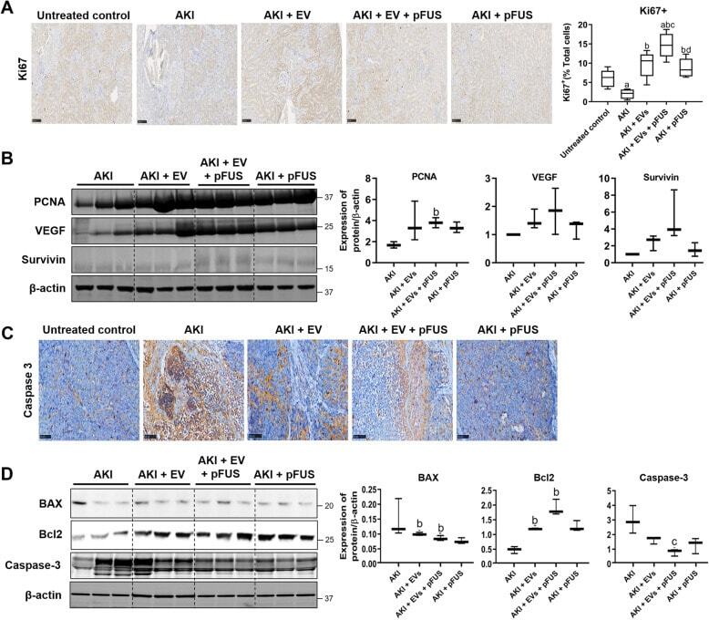
- Fig. 5 Proliferation and apoptosis. a Immunohistochemical staining for proliferation marker Ki67 in kidney tissue (left), alongside quantification of the percentage of Ki67-positive cells (right). b Western blot on kidney tissue measuring proliferation markers PCNA, VEGF, survivin, and beta-actin (left) alongside quantification (right). c Immunohistochemical staining for apoptosis marker Caspase-3 in kidney tissue. d Western blot on kidney tissue measuring apoptosis markers BAX, Bcl-2, and Caspase-3 (left) alongside quantification (right). Each group has n = 3 mice. Significant difference a p < 0.05: relative to untreated control; b p < 0.05: relative to AKI; c p < 0.05: relative to AKI-EV; d p < 0.05: relative to AKI-EV-pFUS