Explore
 Validate
 Learn
Learn Western blot
 Immunocytochemistry
Antibody data
- Antibody Data
- Antigen structure
- References [8]
- Comments [0]
- Validations
- Western blot [2]
- Immunohistochemistry [2]
Submit
Validation data
Reference
Comment
Report error
- Product number
- NB100-56709 - Provider product page

- Provider
- Novus Biologicals
- Proper citation
- Novus Cat#NB100-56709, RRID:AB_837851
- Product name
- Mouse Monoclonal Caspase-3 Antibody
- Antibody type
- Monoclonal
- Description
- Protein G purified.
- Reactivity
- Human
- Host
- Mouse
- Isotype
- IgG
- Vial size
- 0.1 mg
- Concentration
- 1.0 mg/ml
- Storage
- Store at 4C short term. Aliquot and store at -20C long term. Avoid freeze-thaw cycles.
Submitted references Exploiting mitochondrial and oxidative vulnerabilities with a synthetic analog of pancratistatin in combination with piperlongumine for cancer therapy.
Cymbopogon citratus and Camellia sinensis extracts selectively induce apoptosis in cancer cells and reduce growth of lymphoma xenografts in vivo.
Selective Targeting of Cancer Cells by Oxidative Vulnerabilities with Novel Curcumin Analogs.
Terfenadine induces anti-proliferative and apoptotic activities in human hormone-refractory prostate cancer through histamine receptor-independent Mcl-1 cleavage and Bak up-regulation.
Inhibition of HSP27 alone or in combination with pAKT inhibition as therapeutic approaches to target SPARC-induced glioma cell survival.
WRC-213, an l-methionine-conjugated mitoxantrone derivative, displays anticancer activity with reduced cardiotoxicity and drug resistance: identification of topoisomerase II inhibition and apoptotic machinery in prostate cancers.
TNF-alpha renders human peritoneal mesothelial cells sensitive to anti-Fas antibody-induced apoptosis.
TNF-alpha renders human peritoneal mesothelial cells sensitive to anti-Fas antibody-induced apoptosis.
Ma D, Gilbert T, Pignanelli C, Tarade D, Noel M, Mansour F, Gupta M, Ma S, Ropat J, Curran C, Vshyvenko S, Hudlicky T, Pandey S
FASEB journal : official publication of the Federation of American Societies for Experimental Biology 2018 Jan;32(1):417-430
FASEB journal : official publication of the Federation of American Societies for Experimental Biology 2018 Jan;32(1):417-430
Cymbopogon citratus and Camellia sinensis extracts selectively induce apoptosis in cancer cells and reduce growth of lymphoma xenografts in vivo.
Philion C, Ma D, Ruvinov I, Mansour F, Pignanelli C, Noel M, Saleem A, Arnason J, Rodrigues M, Singh I, Ropat J, Pandey S
Oncotarget 2017 Dec 19;8(67):110756-110773
Oncotarget 2017 Dec 19;8(67):110756-110773
Selective Targeting of Cancer Cells by Oxidative Vulnerabilities with Novel Curcumin Analogs.
Pignanelli C, Ma D, Noel M, Ropat J, Mansour F, Curran C, Pupulin S, Larocque K, Wu J, Liang G, Wang Y, Pandey S
Scientific reports 2017 Apr 24;7(1):1105
Scientific reports 2017 Apr 24;7(1):1105
Terfenadine induces anti-proliferative and apoptotic activities in human hormone-refractory prostate cancer through histamine receptor-independent Mcl-1 cleavage and Bak up-regulation.
Wang WT, Chen YH, Hsu JL, Leu WJ, Yu CC, Chan SH, Ho YF, Hsu LC, Guh JH
Naunyn-Schmiedeberg's archives of pharmacology 2014 Jan;387(1):33-45
Naunyn-Schmiedeberg's archives of pharmacology 2014 Jan;387(1):33-45
Inhibition of HSP27 alone or in combination with pAKT inhibition as therapeutic approaches to target SPARC-induced glioma cell survival.
Schultz CR, Golembieski WA, King DA, Brown SL, Brodie C, Rempel SA
Molecular cancer 2012 Apr 5;11:20
Molecular cancer 2012 Apr 5;11:20
WRC-213, an l-methionine-conjugated mitoxantrone derivative, displays anticancer activity with reduced cardiotoxicity and drug resistance: identification of topoisomerase II inhibition and apoptotic machinery in prostate cancers.
Hsiao CJ, Li TK, Chan YL, Hsin LW, Liao CH, Lee CH, Lyu PC, Guh JH
Biochemical pharmacology 2008 Feb 15;75(4):847-56
Biochemical pharmacology 2008 Feb 15;75(4):847-56
TNF-alpha renders human peritoneal mesothelial cells sensitive to anti-Fas antibody-induced apoptosis.
Chen JY, Chi CW, Chen HL, Wan CP, Yang WC, Yang AH
Nephrology, dialysis, transplantation : official publication of the European Dialysis and Transplant Association - European Renal Association 2003 Sep;18(9):1741-7
Nephrology, dialysis, transplantation : official publication of the European Dialysis and Transplant Association - European Renal Association 2003 Sep;18(9):1741-7
No comments: Submit comment
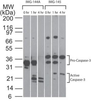
Supportive validation
- Submitted by
- Novus Biologicals (provider)
- Main image
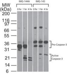
- Experimental details
- Western Blot: Caspase-3 Antibody (31A893) [NB100-56709] - Analysis of Caspase-3 in Jurkat cells. Cells were treated with 2 uM staurosporine for different time periods. Caspase-3 activation is detected in Western blots by the presence of Caspase-3 cleavage fragments. These antibodies detect both pro (full-length) and active (cleaved) Caspase-3, depending on the treatment time points. A basal level of endogenous active Caspase-3 may be see in untreated Jurkat cells.
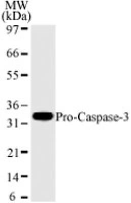
- Submitted by
- Novus Biologicals (provider)
- Main image

- Experimental details
- Western Blot: Caspase-3 Antibody (31A893) [NB100-56709] - Analysis for human Caspase-3 using HL60 lysates with Caspase-3 Antibody (31A893) at 2 ug/mL dilution. NB100-56709 only detects a 32 kD Caspase-3 corresponding to pro-Caspase-3.
Supportive validation
- Submitted by
- Novus Biologicals (provider)
- Main image

- Experimental details
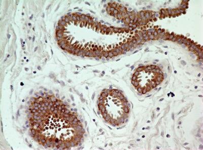
- Immunohistochemistry-Paraffin: Caspase-3 Antibody (31A893) [NB100-56709] - Tissue section of normal human breast using 5 ug/mL concentration of Caspase-3 Antibody (31A893). Very strong diffused as well as granular immunopositivity of Caspase 3 was observed specifically in the cytoplasmic of ductal /acinar epithelial cells.
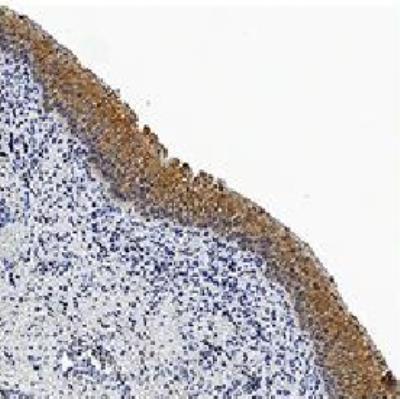
- Submitted by
- Novus Biologicals (provider)
- Main image

- Experimental details
- Immunohistochemistry-Paraffin: Caspase-3 Antibody (31A893) [NB100-56709] - Caspase-3 was detected in immersion fixed paraffin-embedded sections of human bladder tissue using 5 ug/mL of mouse monoclonal Caspase-3 Antibody (31A893) (NB100-56709, Novus Biologicals), for 1 hour at room temperature followed by anti-mouse IgG VisUCyte HRP polymer (VC001). Tissue was stained using DAB (brown) and counterstained with hematoxylin (blue).