Explore
 Validate
 Learn
Learn Western blot
 Immunocytochemistry
Antibody data
- Antibody Data
- Antigen structure
- References [9]
- Comments [0]
- Validations
- Immunocytochemistry [1]
- Immunohistochemistry [1]
Submit
Validation data
Reference
Comment
Report error
- Product number
- HPA002643 - Provider product page

- Provider
- Atlas Antibodies
- Proper citation
- Atlas Antibodies Cat#HPA002643, RRID:AB_1846048
- Product name
- Anti-CASP3
- Antibody type
- Polyclonal
- Description
- Polyclonal Antibody against Human CASP3, Gene description: caspase 3, apoptosis-related cysteine peptidase, Alternative Gene Names: apopain, CPP32, CPP32B, Yama, Validated applications: ICC, IHC, WB, Uniprot ID: P42574, Storage: Store at +4°C for short term storage. Long time storage is recommended at -20°C.
- Reactivity
- Human
- Host
- Rabbit
- Conjugate
- Unconjugated
- Isotype
- IgG
- Vial size
- 100 µl
- Concentration
- 0.1 mg/ml
- Storage
- Store at +4°C for short term storage. Long time storage is recommended at -20°C.
- Handling
- The antibody solution should be gently mixed before use.
Submitted references Comparative pathology and immunohistochemistry of Newcastle disease in domestic chicken (Gallus-gallus domesticus) and Alabio duck (Anas platyrhynchos Borneo)
The appearance of phagocytic microglia in the postnatal brain of Niemann Pick type C mice is developmentally regulated and underscores shortfalls in fine odor discrimination
Proteome study of cutaneous lupus erythematosus (CLE) and dermatomyositis skin lesions reveals IL-16 is differentially upregulated in CLE
Ganglion cells apoptosis in diabetic rats as early prediction of glaucoma: a study of Brn3b gene expression and association with change of quantity of NO, caspase-3, NF-κB, and TNF-α
A High-throughput Bead-based Affinity Assay Enables Analysis of Genital Protein Signatures in Women At Risk of HIV Infection
Auditory cortex interneuron development requires cadherins operating hair-cell mechanoelectrical transduction
Low levels of Caspase-3 predict favourable response to 5FU-based chemotherapy in advanced colorectal cancer: Caspase-3 inhibition as a therapeutic approach.
Connexin 43 and Its Hemichannels Mediate Hypoxia–Ischemia-Induced Cell Death in Neonatal Rats
Photoreceptor damage induced by low-intensity light: model of retinal degeneration in mammals.
Etriwati E, Agungpriyono D, Setiyaningsih S, Darniati D, Ak D, Erwin E, Handharyani E
Open Veterinary Journal 2023;13(4):433
Open Veterinary Journal 2023;13(4):433
The appearance of phagocytic microglia in the postnatal brain of Niemann Pick type C mice is developmentally regulated and underscores shortfalls in fine odor discrimination
Rava A, La Rosa P, Palladino G, Dragotto J, Totaro A, Tiberi J, Canterini S, Oddi S, Fiorenza M
Journal of Cellular Physiology 2022;237(12):4563-4579
Journal of Cellular Physiology 2022;237(12):4563-4579
Proteome study of cutaneous lupus erythematosus (CLE) and dermatomyositis skin lesions reveals IL-16 is differentially upregulated in CLE
Niewold T, Meves A, Lehman J, Popovic-Silwerfeldt K, Häyry A, Söderlund-Matell T, Charlesworth C, Madden B, Lundberg I, Wahren-Herlenius M, Svenungsson E, Oke V
Arthritis Research & Therapy 2021;23(1)
Arthritis Research & Therapy 2021;23(1)
Ganglion cells apoptosis in diabetic rats as early prediction of glaucoma: a study of Brn3b gene expression and association with change of quantity of NO, caspase-3, NF-κB, and TNF-α
Tjandra I, Artini W, Siregar N, Victor A
International Journal of Ophthalmology 2020;13(12):1872-1879
International Journal of Ophthalmology 2020;13(12):1872-1879
A High-throughput Bead-based Affinity Assay Enables Analysis of Genital Protein Signatures in Women At Risk of HIV Infection
Månberg A, Bradley F, Qundos U, Guthrie B, Birse K, Noël-Romas L, Lindskog C, Bosire R, Kiarie J, Farquhar C, Burgener A, Nilsson P, Broliden K
Molecular & Cellular Proteomics 2019;18(3):461-476
Molecular & Cellular Proteomics 2019;18(3):461-476
Auditory cortex interneuron development requires cadherins operating hair-cell mechanoelectrical transduction
Libé-Philippot B, Michel V, Boutet de Monvel J, Le Gal S, Dupont T, Avan P, Métin C, Michalski N, Petit C
Proceedings of the National Academy of Sciences 2017;114(30):7765-7774
Proceedings of the National Academy of Sciences 2017;114(30):7765-7774
Low levels of Caspase-3 predict favourable response to 5FU-based chemotherapy in advanced colorectal cancer: Caspase-3 inhibition as a therapeutic approach.
Flanagan L, Meyer M, Fay J, Curry S, Bacon O, Duessmann H, John K, Boland KC, McNamara DA, Kay EW, Bantel H, Schulze-Bergkamen H, Prehn JH
Cell death & disease 2016 Feb 4;7(2):e2087
Cell death & disease 2016 Feb 4;7(2):e2087
Connexin 43 and Its Hemichannels Mediate Hypoxia–Ischemia-Induced Cell Death in Neonatal Rats
Wang J, Ma A, Xi J, Wang Y, Zhao B
Child Neurology Open 2014;1(1)
Child Neurology Open 2014;1(1)
Photoreceptor damage induced by low-intensity light: model of retinal degeneration in mammals.
Contín MA, Arietti MM, Benedetto MM, Bussi C, Guido ME
Molecular vision 2013;19:1614-25
Molecular vision 2013;19:1614-25
No comments: Submit comment
Supportive validation
- Submitted by
- Atlas Antibodies (provider)
- Main image

- Experimental details
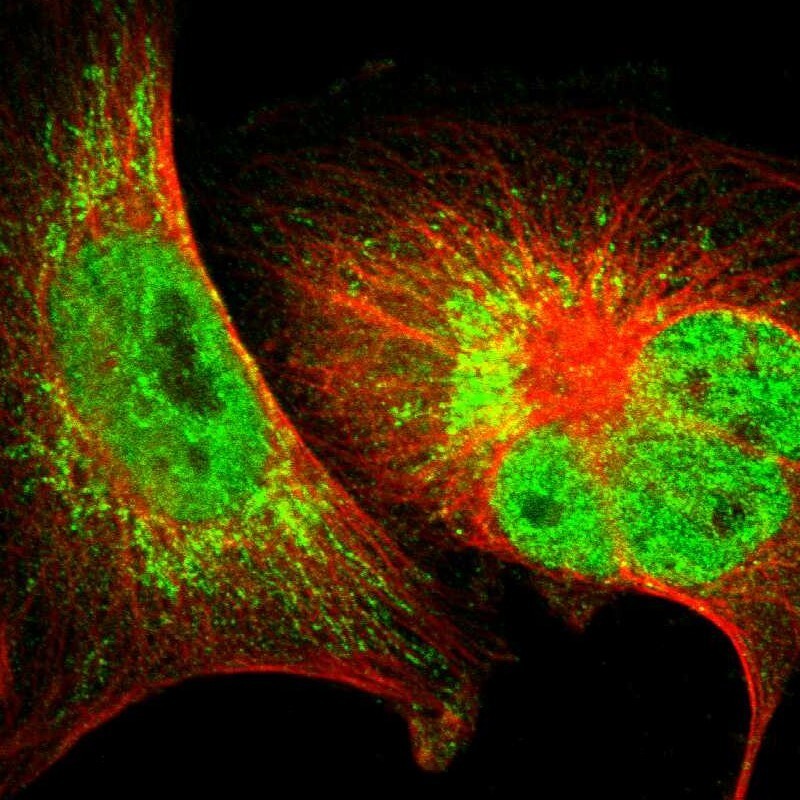
- Immunofluorescent staining of human cell line U-251 MG shows localization to nucleoplasm & mitochondria.
- Sample type
- Human
Supportive validation
- Submitted by
- Atlas Antibodies (provider)
- Enhanced method
- Orthogonal validation
- Main image

- Experimental details
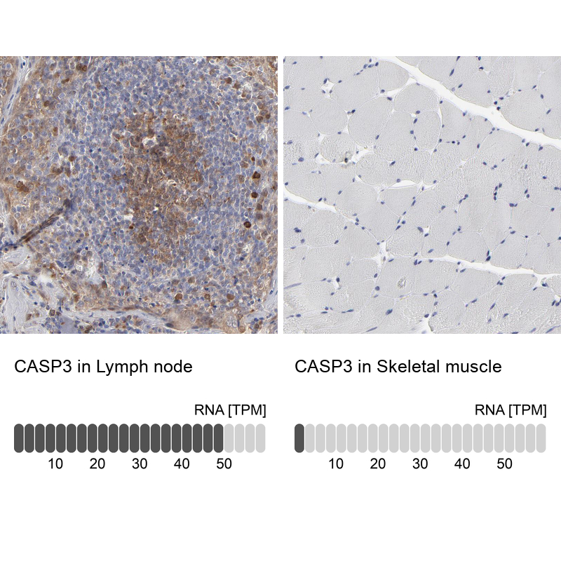
- Immunohistochemistry analysis in human lymph node and skeletal muscle tissues using HPA002643 antibody. Corresponding CASP3 RNA-seq data are presented for the same tissues.
- Sample type
- Human
- Protocol
- Protocol